Categories
Case Reports

Continuous positive airway pressure for obstructive sleep apnoea improved oculogyric crises as well as psychotic symptoms in a woman with schizophrenia and developmental disability

Abstract

Introduction: This report highlights the risk factors and complexities of schizophrenia as well as the adverse effects of treatment. Obstructive sleep apnoea (OSA) has a notorious history of under-diagnosis in both the general population as well as those suffering from mental health disorders, particularly schizophrenia. Antipsychotics have life altering side effects contributing both to a decrease in quality of life as well as increasing morbidity and mortality.

Case overview: This case report presents a 61-year-old female with diagnoses of schizophrenia, frontal lobe epilepsy, a developmental disability, oculogyric crises (OGC), and obstructive sleep apnoea.

Discussion overview: Early intervention with continuous positive airway pressure (CPAP) in those suffering from OSA can have dramatic effects decreasing the burden of concurrent disease. This report showcases that treatment of OSA with CPAP increased patient wellbeing, allowing down-titration of risperidone, and thereby ameliorating the drug-induced OGC in this patient. 

Download (PDF, 148KB)

Categories
Case Reports

A patient with right striatocapsular stroke complicated by relative adrenal insufficiency

Introduction: Relative adrenal insufficiency can occur throughout the progression of critical illness and is generally transient.

Case: This case report describes a 74 year-old male with right hemispheric stroke syndrome on a background of multiple cardiovascular risk factors. A CT scan showed no acute change.
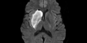
An MRI scan revealed an acute right striatocapsular infarction. No acute therapies (thrombolysis or endovascular clot retrieval) were performed, as the time of symptom onset was unknown (patient awoke with symptoms). One week later, hyponatraemia was noted with a concurrent decline in function. A repeat MRI showed no interval change or haemorrhagic transformation to account for the functional decline. Complications included relative adrenal insufficiency, diagnosed presumptively and managed with cortisone, and gait instability managed with rehabilitation and allied health input.

Discussion: We review the literature concerning the association between acute ischaemic stroke and adrenal insufficiency and the clinical and biochemical overlap in our patient. This case report aims to increase awareness of relative adrenal insufficiency following a stroke and provide a discussion of possible mechanisms.

Download (PDF, 1.65MB)

Categories
Case Reports

The rare case of a pelvic abscess following caesarean section in a Sri Lankan woman: an argument for medical student electives

Abstract

Introduction: A pelvic abscess is a rare complication that can occur following gynaecological and obstetric procedures. Whilst the condition is not confined geographically, women in less developed countries are at an increased risk of developing this complication, due in part to low resource settings, socioeconomic status, and educational attainment.

Case: A 36 year old primigravid woman in rural Sri Lanka undergoing a non-emergency caesarean section delivery developed clinical signs of puerperal sepsis two days postpartum. Following transfer to a tertiary hospital, imaging and laparotomy confirmed the presence of a pelvic abscess associated with the caesarean section wound. Surgical drainage was performed and IV antibiotics were administered, ultimately resulting in the full recovery of the patient. The infant did not demonstrate clinical signs of sepsis at birth.

Discussion: Numerous factors contributed to the development of this serious complication in this patient, including increased maternal age at first pregnancy, caesarean section management, and the low resource setting of the patient’s care. This case also highlights the difficulties and barriers facing patients and doctors in less developed countries, including that of patient transfers from a rural setting and the availability of specific antibiotics recommended in clinical guidelines. The barriers to optimal care faced by this woman are largely disconnected from the experiences of patients that Australian medical students see in their day-to-day training. Overseas electives to low resource areas should be required and supported amongst all Australian medical students to facilitate greater appreciation for such barriers and to foster their skills in resourcefulness and empathy.


Introduction

As a demonstration of the public health issues and associated complications that arise in less developed countries, the following presents a rare case of pelvic abscess formation following caesarean section delivery. This is written from the perspective of an Australian medical student on an obstetrics and gynaecology elective in rural Sri Lanka.

Pelvic abscess is considered a rare complication of pelvic surgery, affecting less than 1% of women undergoing any obstetric or gynaecological procedure [1]. The implications of such a complication are more significant when they occur in a low resource setting, such as Sri Lanka [2]. Data suggest puerperal sepsis accounts for 11.6% of maternal mortality in such settings, compared with 2.1% in developed countries [3]. A range of factors contribute to the risk of puerperal sepsis and pelvic abscess, including increasing maternal age at first pregnancy, high caeserean section rates, and low socioeconomic and educational status, all well-documented factors present in this patient and their demographic [2,4,5]. Whilst pelvic abscess is a complication that has no geographical boundaries, women in less developed countries are at increased risk of dying from this complication [3]. This is likely compounded by sub-optimal infection control and limited access to resources, including trained midwifery and obstetric care [2]. This is reflected in the high rate of hospital-acquired infections, up to 50%, resulting from surgical site wounds [3]. Despite the high morbidity and mortality of pelvic abscesses and puerperal sepsis, the actual incidence of both conditions is poorly defined [3]. The following case exemplifies some of these challenges.


Case

A 36 year old primigravid woman underwent a caesarean section with no immediate surgical complications, in the setting of a reportedly uneventful pregnancy. Surgery was performed at a local rural hospital in Sri Lanka, approximately three to four hours by road transfer from the capital, Colombo. The reason for caesarean section was unclear; however, it was understood that the caesarean section was performed in a non-emergency setting. Two days postpartum, the woman developed fever (temperature not specified) and complained of a triad of foul-smelling and purulent lochia, abdominal distension, and dyspnoea, according to the patient and her family. The patient was denied transfer by the treating hospital, and so the family arranged private transportation to a tertiary centre in Colombo. She arrived in a state of septic shock: mildly hypothermic (36.0˚C), tachypnoeic (45 breaths/min), hypertensive (150/100 mmHg), tachycardic (150 beats/min), and cyanotic with significant generalised oedema. Computerised tomography of the abdomen and pelvis identified abscess formation anterior to the uterine sutures and significant fluid in the peritoneal cavity. An emergency laparotomy was performed. A dehiscence of her uterine scar was repaired and two litres of pus was drained. The fluid cultured positive for Group B beta-haemolytic Streptococcus, sensitive to vancomycin. The patient was subsequently commenced on intravenous antibiotics (vancomycin, metronidazole, and meropenem) post-operatively.

Following surgery, hyperglycaemia (180-250 mg/dL) and hypertension (160/110 mmHg) persisted for some weeks, with recovery complicated by a secondary wound infection requiring additional antibiotics (flucloxacillin and metronidazole) and a second laparotomy. Mother and baby ultimately recovered completely, with no clinical signs of postnatal sepsis reported in the child. Clinical guidelines for puerperal sepsis recommend a combination of broad-spectrum antibiotics, targeting the common polymicrobial sources, including anaerobic organisms (E. coli and S. pyogenes), and surgical abscess drainage [5,6]. This case failed to utilise the recommended first-line empirical antibiotics. Poor antibiotic practices, unreliable antibiotic supplies, and a lack of adequate medication delivery protocols are cited as common reasons for increasing rates of puerperal sepsis in the developing world, with one or more of these factors potentially influencing antibiotic choice in this case [2,4].


Discussion

Although the woman had a full recovery, this case serves as a reminder of the seriousness of obstetric complications, and how they may be compounded by geographical factors and a paucity of adequate medical resources. This case offered significant opportunity for learning and personal reflection. Primarily, I was able to recognise that the outcomes of childbirth and postnatal care in less developed countries may be vastly different to such events occurring in Australia. Some of the difficulties I observed included inadequate systems for medical documentation, and communication of results and information for clinical handover. The disparity in access to such basic resources is alarming, and I believe all medical professionals should be angry about such injustices. To fully understand this degree of inequality, however, it required the immersive experience of an elective term in Sri Lanka. Based on my own experience, I would strongly recommend an overseas elective as a mandatory component of any Australian medical degree. This could be a useful step in helping Australian graduates to become more resourceful. In addition, it will help our graduates recognise the importance of an effective and well-resourced, high-quality health system in achieving the best health outcomes for our patients.


Consent declaration
Informed consent was obtained from the patient for publication of this case report.

Acknowledgements
Dr Roshan Zaid (consultant obstetrician and gynaecologist, Nawaloka Hospital, Colombo, Sri Lanka), a senior consultant involved in the above patient’s care, who provided clinical advice and support in compiling this report.

Financial assistance for this elective was provided in the form of a bursary from the University of Melbourne.

Conflict of interest
None declared.


References

  1. Mahdi H, Goodrich S, Lockhart D, DeBernardo R, Moslemi-Kebria M. Predictors of surgical site infection in women undergoing hysterectomy for benign gynecologic disease: a multicenter analysis using the national surgical quality improvement data. J Minim Invasive Gynecol. 2014;21(5):901-9.
  2. Buddeberg BS, Aveling W. Puerperal sepsis in the 21st century: progress, new challenges and the situation worldwide. Postgrad Med J. 2015;91(1080):572-8
  3. Hussein J, Walker L. Puerperal sepsis in low- and middle-income settings: past, present and future. In: Kehoe S, Neilson J, Norman J, editors. Maternal and infant deaths – chasing millennium development goals 4 and 5. Cambridge: Cambridge University Press; 2010.
  4. Knowles S, O’sullivan N, Meenan A, Hanniffy R, Robson M. Maternal sepsis incidence, aetiology and outcome for mother and fetus: a prospective study. BJOG. 2015;122(5):663-71.
  5. The World Health Organisation. Managing puerperal sepsis – midwifery education module 4: avoidable factors. 2nd ed. Geneva: The WHO Press; 2008.
  6. The Royal College of Obstetrics and Gynaecology. Bacterial sepsis following pregnancy. Green-top Guideline No. 64B [Internet]. 2012 [cited 2016 Nov]. Available from: https://www.rcog.org.uk/en/guidelines-research-services/guidelines/gtg64b/
Categories
Case Reports

An unusual aetiology in a patient with increasing abdominal girth

Pseudomyxoma peritonei (PMP) is a rare, slow growing mucinous ascites, typically associated with primary appendiceal or ovarian neoplasm [1]. Mucinous material fills the peritoneal cavity, causing enlargement of the abdomen and has been described as “jelly belly”, due to its appearance at laparotomy [2]. The symptoms of PMP are often non-specific and vague, causing difficulties in diagnosis. Further, diagnostic imaging is not always able to detect the disease prior to surgery. The clinical implications of this are that PMP is not commonly considered a differential diagnosis in patients with these symptoms, which may then delay the diagnosis being made. This causes a potential delay in treatment, which has been shown to worsen the morbidity and mortality associated with PMP [3,4].

Introduction

Pseudomyxoma peritonei (PMP) is a rare, slow growing mucinous ascites, typically associated with primary appendiceal or ovarian neoplasm [1]. Mucinous material fills the peritoneal cavity, causing enlargement of the abdomen and has been described as “jelly belly”, due to its appearance at laparotomy [2]. The symptoms of PMP are often non-specific and vague, causing difficulties in diagnosis. Further, diagnostic imaging is not always able to detect the disease prior to surgery. The clinical implications of this are that PMP is not commonly considered a differential diagnosis in patients with these symptoms, which may then delay the diagnosis being made. This causes a potential delay in treatment, which has been shown to worsen the morbidity and mortality associated with PMP [3,4].

Case

A 48-year-old female presented to her general practitioner with a two-month history of increasing abdominal girth and a feeling of pelvic “fullness”. Importantly, she had not been unwell, did not have any infective symptoms, no loss of weight or appetite, and no nausea or vomiting. Her bladder and bowel function was normal, her periods were regular, and her Pap smears were up to date and normal.

The patient had a past medical history of primary hypothyroidism and depression, both of which were clinically stable. She had no known allergies. Her regular medications were Fluoxetine (20 mg daily) and thyroxine sodium (100 mg daily). She lived with her 16-year-old daughter, and worked full time in a delicatessen. Her family history included bowel, prostate, and breast cancer.

Her GP ordered various investigations (Table 1). Her borderline high CA-125 level (38 kU/L, reference range < 36 kU/L) and the imaging findings suggested a possible gynaecological malignancy.

Table 1: Initial investigation results

Full blood examination Normal
UEC Normal
Ca125^ 38 (RR* < 36 kU/L)
CEA^ 7.2 (RR* < 2.5 mg/L in non-smokers)
Ca19.9^ 12 (RR* < 31 kU/L)
Trans-abdominal ultrasound
  • Normal-sized, anteverted uterus.
  • Right adnexa: large complex-appearing mass lesion associated with ascites. Right ovary not able to be visualised.
  • Left adnexa: left ovary slightly bulky but unremarkable.
CT chest/ abdomen/ pelvis Relevant features:

  • Multiloculated cystic lesion noted within the right side of the pelvis that measures 9 cm in maximum diameter.
  • Thin septa with associated calcifications are noted.
  • Ascites is present in the peritoneal cavity and there is streaking of the omentum. Some of this streaking is suspicious for omental seeding.
  • There is no retroperitoneal lymphadenopathy seen

Conclusion: right-sided pelvic lesion consistent with ovarian mucinous cystadenomatous-type lesion. The presence of ascites and possible omental caking suggests adenocarcinoma.

  • *RR: reference range
  • ^Ca125: cancer antigen 125, CEA: carcinoembryonic antigen, Ca19.9: cancer antigen 19.9. These are common tumour markers used in conjunction with clinical examination and other investigations to aid cancer diagnosis.

She was referred to an outpatient gynaecological oncology clinic for further evaluation and formulation of a management plan. On examination in the clinic, the patient looked well and was afebrile. Her abdomen was distended, with a palpable, non-tender mass in the right iliac fossa. Mild ascites was present. She was also obese (BMI 37). Per vaginal examination revealed a palpable mass, with noted fixation of the right adnexa. Her uterus was mobile, non-tender, and of normal size and morphology.

The patient was discussed at the multidisciplinary team meeting where it was recommended that she undergo a laparotomy for total abdominal hysterectomy, bilateral salpingo-oophrectomy, and omentectomy. At the time of the surgery, she was noted to have extensive mucinous material throughout her peritoneum, and within her uterus and cervix. The mass seen on imaging was found to be an enlarged appendix, which required concurrent general surgical consultation for removal. The specimens were sent to pathology for analysis (Table 2).

Table 2: Formal pathology results

Cytology
  • Smears are mildly cellular, with abundant thick mucin, in keeping with peritoneal mucinosis or pseudomyxoma peritonei.
  • No overt malignant cells identified.
Histopathology Macroscopic
  • Uterus, tubes and ovaries: serosal surface of the uterus appears haemorrhagic, with multiple mucin-containing cystic lesions. No mass lesion is identified. The left ovary is haemorrhagic with a disrupted cyst present. The right ovary is partially covered in mucinous material and cysts. Unremarkable myometrium.
  • Appendix: large specimen (9 cm x 6 cm x 5 cm) that is disrupted, cystically-dilated and containing mucin. The surface is congested, with mucin extravasation present.
Microscopic
  • Uterus, tubes and ovaries: all sections of the serosa have organising mucinous exudate present. No tumour cells are seen in the serosal mucous. No neoplasia is seen in either ovary.
  • Appendix: dilatation of the proximal lumen, with marked fibrosis and patchy calcifications. Distally the lumen is distended by mucin, with tall columnar epithelium showing basophilic hyperchromatic nuclei and suprabasaloid mucin production. Some of these atypical cells are spread over the surface of the luminal mucus. The mucin has dissected through the appendix muscularis to rupture into the peritoneal cavity.

The subsequent histopathological diagnosis was of a primary appendiceal malignancy, with rupture and extensive mucin extrusion into the peritoneal cavity.

She had an unremarkable post-operative course, and was discharged home on day 4. She was to be followed up with the pathology results for relevant discussion regarding her ongoing treatment, management, and prognosis.

Discussion

Pathology

The underlying pathology in PMP has been a controversial area for some time [6]. The pathological process was originally thought to be due to a foreign body reaction after mucus containing cysts ruptured into the peritoneum [7]. However, it has now been re-defined to embrace a spectrum of cells from benign to malignant that produce abundant mucinous fluid. Within the ascitic fluid, there may be a few, if any, neoplastic cells seen, as the mucinous exudate is believed to spread further than any potential malignant cells within the peritoneum [8]. Malignant cells that produce PMP are often described as histologically borderline, as they do not show invasion of surrounding structures since they adhere rather than invade. Haematogenous or lymphatic metastasis is unusual, and most cases are found to originate from the appendix, with the most common being primary appendiceal mucinous neoplasia [6]. Rarely, however, the origin may be from the ovary, stomach, gallbladder, pancreas, urinary bladder, uterus, or fallopian tubes [1]. The mucinous tumour cells form cysts that increase intraluminal pressure within the organ of origin, and eventually cause the luminal wall to rupture [8]. The cells are then able to leak into the peritoneum. They are transported passively by peritoneal fluid flow and absorption, and by gravity to adhere to both abdominal and pelvic structures. Even if PMP is of a benign cell origin, the slow but relentless increase of gelatinous fluid in the peritoneal cavity causes compression of intra-abdominal organs, and mechanical and functional gastrointestinal obstruction [8].

Clinical presentation

Symptoms of PMP vary and will depend on the extent of the disease. Most commonly, patients report increasing abdominal girth or enlarging incisional, umbilical, or inguinal hernias [2]. Women may be diagnosed incidentally during routine pelvic examination or may present with infertility [2]. Patients may also report early satiety, as the space within the peritoneal cavity for the stomach to expand decreases, or with a clinical picture of acute appendicitis [9]. However, PMP is still often diagnosed incidentally at laparotomy, with symptoms sometimes inaccurately labeled as irritable bowel syndrome for years prior to diagnosis [1,2].

Utility of diagnostic imaging in PMP

Multiple imaging modalities have been reviewed with regard to PMP. Plain abdominal x-rays have been found to be of little diagnostic use, however, it may help to diagnose intestinal obstruction, a late complication of PMP [10]. Ultrasound may be utilised, with reported findings including homogenous tumour deposits, separated ascites, scalloping of the liver edges, and echogenic masses [11]. CT scans are the most widely used imaging technique for intra-abdominal pathology. Findings suggestive of PMP include scalloping of organs, ascitic septations and loculi, curvilinear calcifications, and omental thickening [10].

A review of CT scan use in 17 cases of PMP reported that early disease is easier to diagnose than more advanced disease. The authors urged radiologists to look for a pattern of mucinous ascites accumulation, rather than the appearance of individual deposits of disease on the image [12]. These authors were based at a surgical hospital and had experience with PMP. It may be difficult to expect a radiologist to detect this diagnosis without having had a similar level of experience. Ultrasound requires similar expertise, where it has been reported that familiarity with the features of PMP are required for accurate diagnosis [13].

Clinical implications

Despite being uncommon, PMP is a possible diagnosis that may occur in patients. It is worth keeping this disease as a differential diagnosis for patients that present with abdominal fullness. Imaging may help with the diagnosis but is not definitive. Without treatment, the prognosis for this condition is poor, with a ten-year survival rate of approximately 32% [14]. Treatments such as peritonectomy, intra-peritoneal chemotherapy at the time of surgery, and radical de-bulking of tumour deposits have been shown to improve the recurrence free survival time in these patients and decrease overall mortality [3,4]. Further, surgery that does not definitively de-bulk the condition contributes to increased difficulty in managing PMP effectively later on, through the creation of adhesions that can facilitate spread of PMP to the small bowel [3]. Early diagnosis is therefore important to help expedite care, allow for appropriate surgical and oncology management to occur, and improve outcomes in patients with PMP.

This case highlights that although it is most commonly horses when you hear hooves, very occasionally, it may actually be a zebra.

Consent declaration

Informed consent was obtained from the patient for publication of this case report.

Conflicts of interest

None declared.

References

[1] Sherer DM, Abulafia O, Eliakim R. Pseudomyxoma Peritonei: a review of current literature. Gynecol Obstet Invest. 2001;51:73-80.

[2] Brueggan C, Baird G, Meisheid A. Pseudomyxoma peritonei syndrome of appendiceal origin: an overview. Clin J Onc Nursing. 2007;11(4):525-532.

[3] Sugarbaker PH. New standard of care for appendiceal epithelial neoplasms and pseudomyxoma peritonei syndrome. Lancet Oncol. 2006;7(1):69-76.

[4] Yan TD, Black D, Savady R, Sugarbaker PH. Systematic review on the efficacy of cytoreductive surgery and perioperative intraperitoneal chemotherapy for pseudomyxome peritonei. Ann Surg Oncol. 2007;14(2):484-492.

[5] Mann WJ, Wagner J, Cumas J, Chalas E. The management of pseudomyxoma peritonei. Cancer. 1990;66:1636-40.

[6] Carr NJ, Finch J, Ilesley IC, Chandrakumaran K, Mohamed F, Mirnezami A, et al. Pathology and prognosis in pseudomyxoma peritonei: a review of 274 cases. J Clin Pathol. 2012;65:919-923.

[7] Jivan S, Bahal V. Pseudomyxoma peritonei. Postgrad Med J. 2002;78:170-2.

[8] Agrawal AK, Bobiński P, Grzebieniak Z, Rudnicki J, Marek G, Kobielak P, et al. Pseudomyxoma peritonei originating from the urachus – case report and review of the literature. Curr Oncol. 2015 Feb;21(1):e155-165.

[9] Esquivel J, Sugarbaker PH. Clinical presentation of the pseudomyxoma peritonei syndrome. Br J Surg. 2000; 87:1414-18.

[10] Walensky RP, Venbrux AC. Prescott CA, Osterman FA Jr. Pseudomyxoma peritonei. Am J Roentgenol. 1996;167:471-4.

[11] Seshul MB, Coulam CM. Pseudomyxoma peritonei: computed tomography and sonography. Am J Roentgenol. 1981;136:803-6.

[12] Sulkin TV, O’Neill H, Amin AI, Moran B. CT in pseudomyxoma peritonei: a review of 17 cases. Clin Radiol. 2002;57:608-613.

[13] Li Y, Guo A, Tang J, Wang L, Wang J, Yu D. Role of preoperative sonography in the diagnosis and pathologic staging of pseudomyxoma peritonei. J Ultrasound Med. 2013;32(9):1565-1570.

[14] Gough DB, Donohue JH, Schutt AJ, Gonchoroff N, Goellner JR, Wilson TO, et al. Pseudomyxoma peritonei: long term patient survival with an aggressive regional approach. Ann Surg. 1994;219(2):112-119.

Categories
Case Reports

Focal segmental glomerulosclerosis: Treatment beyond corticosteroids

This case report describes a fourteen year-old male who presented with a relapse of steroid-dependent focal segmental glomerulosclerosis (FSGS). FSGS is responsible for 10-15% of cases of idiopathic nephrotic syndrome (INS) in children, with the majority of cases attributed to minimal change disease. Prednisolone is first line for the induction of remission, with the majority of INS cases responding to initial therapy. Those who fail to achieve remission within four weeks of corticosteroid therapy are labeled “steroid-resistant”. Of those who do remit with corticosteroids, 80% have a relapse, with 50% of these patients having “frequently relapsing disease”. Those patients who relapse while on corticosteroids, or within two weeks of cessation of corticosteroids, are labeled “steroid-dependent”. The aim of this article is to review the literature available on the management of FSGS, particularly steroid-resistant, steroid-dependent, and frequently relapsing disease.

Case Study

ML, a fourteen year-old male, presented to a rural paediatric department with a one-month history of increasing oedema of his face, sacrum, and lower limbs; lethargy; and oliguria on a background of known steroid-dependent focal segmental glomerulosclerosis (FSGS).

ML first presented with nephrotic syndrome in late 2014, which was initially responsive to corticosteroids, but relapsed following steroid cessation. A renal biopsy was performed in early 2015 and ML was diagnosed with FSGS. At this time, he was started on cyclosporin 125 mg OD and was managed by a general paediatrician and nephrologist.

Approximately one month prior to his admission, ML commenced 50 mg doxycycline at night for acne and the cyclosporin was consequently reduced to 100 mg daily due to concerns that doxycycline may increase the cyclosporin concentration. Soon after, his symptoms of nephrotic syndrome began to return and the cyclosporin was increased to 110 mg daily. ML had also started ramipril 1.25 mg at night prior to his admission.

ML had a history of partial seizures, diagnosed in 2008, which were well controlled by valproate 400 mg twice daily. Developmental history was unremarkable. He had no known allergies and had received his routine childhood vaccinations. Due to the immunosuppressive nature of relapsing nephrotic syndrome, he also received the pneumococcal vaccine and an annual influenza vaccine. There was a family history of epilepsy in his grandmother, but no family history of renal disease. ML was an only child, a non-smoker, and a non-drinker, who lived with his mother in a major regional centre.

On examination, ML was pale and lethargic, with marked periorbital oedema. His vital signs were within normal limits. He had cold peripheries, indicating intravascular depletion but central capillary refill was normal. His jugular venous pressure (JVP) was not elevated, but he had pitting oedema extending to the upper legs, as well as sacral, periorbital, and scalp oedema. His abdomen was distended and ascites was demonstrated by shifting dullness. The abdomen was otherwise non-tender and bowel sounds were present. Heart sounds were dual with no murmurs. His chest was clear with resonant percussion, excluding pulmonary oedema.

Investigations included urine dipstick; urine microscopy, culture, and sensitivity (MCS); spot protein-creatinine ratio; full blood examination (FBE); urea, electrolytes, and creatinine (UEC); liver function tests (LFTs); and a cyclosporin level. Urinary investigations revealed heavy proteinuria, but no haematuria, and all other investigations were unremarkable.

ML was admitted for management of his acute relapse, which included fluid and salt restriction, daily weighs, and daily urine dipstick. The ramipril was ceased. He was administered 75 mg of intravenous 20% albumin over six hours, with 40 mg of intravenous frusemide given at mid-infusion and post-infusion.

ML lost four kilograms overnight and was discharged on a five-day course of oral frusemide, with a 40 mg dose on the first day, then 20 mg for four days.

Discussion

Background

FSGS is a histopathological pattern of glomerular injury seen under light microscopy, in which sclerosis occurs in segments of only some of the glomeruli [1]. This pattern of injury can occur in all age groups and is the most common cause of adult nephrotic syndrome [2]. FSGS is also identified in 10-15% of cases of idiopathic nephrotic syndrome in children, with the majority of cases attributed to minimal change disease [3].

In most cases of FSGS, the underlying cause is unknown – termed “primary FSGS” [4]. However, secondary FSGS may develop as a response to previous renal injury. Underlying causes of secondary FSGS include reflux nephropathy, infections (for example, HIV), obesity, medications (for example, interferon), genetic mutations, surgical resection of renal tumours, congenital renal dysplasia, and intrauterine growth restriction [5].

Primary FSGS presents with a typical nephrotic syndrome, including foamy urine and extensive oedema [6], particularly periorbital oedema. Nephrotic syndrome is confirmed with a spot urine protein creatinine ratio >0.2 g/mmol [3]. Secondary FSGS is more variable in its presentation, with proteinuria often below nephrotic levels and patients being less likely to present with overt oedema [5].

In children who present with overt nephrotic syndrome, a renal biopsy is not appropriate, because the majority of these cases will be due to minimal change disease. Only when they are unresponsive to corticosteroids, or develop a frequently relapsing or steroid-dependent pattern of disease, is a renal biopsy justified [6]. For indistinct presentations (for example, proteinuria below nephrotic levels), a renal biopsy may be considered on the initial presentation [7]. The risks and benefits of the renal biopsy must be evaluated, with post-biopsy bleeding being a major risk to consider [6].

The following discussion will focus on the treatment of primary FSGS. Secondary FSGS is best treated with angiotensin converting enzyme (ACE) inhibitors or angiotensin receptor blockers to lower the intraglomerular pressure and treatment of the underlying cause, when possible [2].

Immunosuppressive treatment: corticosteroid

Corticosteroids are first-line in treatment of idiopathic nephrotic syndrome (INS) for the induction of remission. Between 80-90% of cases of INS are responsive to initial corticosteroid therapy [3]. Those patients who fail to achieve remission within four weeks of corticosteroid therapy are labeled “steroid-resistant”. Of those patients who respond initially, there is an 80% chance of relapse, with 50% of those having frequently relapsing disease, defined as two or more relapses in the first six months or four or more relapses in any twelve-month period [3,8]. Those who relapse while on corticosteroids or within two weeks of cessation of corticosteroids are labeled “steroid-dependent” [3].

Immunosuppressive treatment: non-corticosteroid

In steroid-resistant, steroid-dependent, and frequently relapsing disease, non-corticosteroid immunosuppressive agents are utilised. The available evidence for each of the commonly used non-corticosteroid immunosuppressive agents will be explored to determine if cyclosporin is the best treatment to prevent relapse in a patient like ML, who has steroid-dependent FSGS.

Calcineurin inhibitors, with or without low dose prednisolone, are first line [1]. The majority of evidence is with cyclosporin. Cochrane Reviews have demonstrated that cyclosporin increases the rate of remission in children with steroid-resistant disease [9] and reduces relapses in steroid-dependent disease, compared with prednisolone alone [8]. Cyclosporin was superior to intravenous cyclophosphamide in steroid-resistant disease [9]. However, in steroid-dependent disease, relapse was reduced with an eight-week course of alkylating agents, cyclophosphamide, or chlorambucil, while cyclosporin required a prolonged course and its effects were not always sustained following treatment cessation [8]. Therefore, cyclophosphamide plays no role in steroid-resistant disease, but may be used in the treatment of steroid-dependent FSGS when cyclosporin has failed or in patients with higher risk of calcineurin nephrotoxicity due to extensive interstitial fibrosis or vascular disease [1].

Mycophenolate motefil may be useful as an alternative medication for relapsing disease, however, the evidence is limited to a few smaller trials [8]. It may be used in combination with corticosteroids when calcineurin inhibitors have been unsuccessful or are contraindicated. Rituximab is another alternative, which has had some success in steroid-dependent disease, but the evidence does not support its use in steroid-resistant disease [8,10,11]. Subcutaneous natural adrenocorticotropic hormone (ACTH) therapy has also had some success in pilot studies, however, the treatment is expensive and further randomised trials are required to confirm the results [12,13].

Non-immunosuppresive treatment

The evidence clearly supports the use of ACE inhibitors or angiotensin receptor blockers in children with steroid-resistant nephrotic syndrome and secondary FSGS [7]. The use of these agents in steroid-dependent or frequently relapsing disease has not been specifically studied. However, guidelines on the use of anti-hypertensive agents in children with chronic kidney disease from any cause suggest that children should be started on an ACE inhibitor or angiotensin receptor blocker when their blood pressure is consistently above the 90th percentile for their age, sex, and height [14]. Treatment should aim to reduce blood pressure to at or below the 50th percentile, unless limited by symptomatic hypotension [14]. Blood pressure-lowering drugs should be used when indicated, irrespective of the level of proteinuria [14]. In primary FSGS, blood pressure-lowering therapy may slow progression to end-stage renal disease, however, it rarely results in remission without concurrent immunosuppressive treatment [15].

Hyperlipidaemia is a common complication of nephrotic syndrome. Combined with the higher cardiovascular risk of patients with chronic kidney disease, this calls for lipid-lowering therapy with a statin [1,16]. While lipid-lowering agents have been successful in lowering lipids in adults with nephrotic syndrome, no studies have looked at the mortality and morbidity benefits of a statin [16]. The use of statins in children with nephrotic syndrome is controversial, with small studies showing that statins reduce lipid levels and are well tolerated, however, there is a lack of evidence regarding long-term safety of statins in paediatric patients [17].

Renal transplantation

Over ten years, 60% of cases of FSGS progress to end-stage renal failure [18]. These patients will need dialysis or renal transplantation. However, there is a high rate of graft failure, with recurrence of FSGS in 30% of allografts [19,20]. The graft survival is lower in children than in adults [19].

Therapeutic plasmapheresis, used for a number of antibody-mediated conditions, is a process that removes the antibody-containing plasma from the patient’s blood and replaces it with unaffected plasma or a plasma substitute [21]. Therapeutic plasmapheresis may be used in FSGS prophylactically before transplantation or in the treatment of established recurrence in an allograft [19]. Studies show that 49-70% of children with recurrent FSGS who receive plasmapheresis enter complete or partial remission of proteinuria [19]. A small study demonstrated that early and intensive daily plasmapheresis in patients with recurrence was beneficial in obtaining complete remission [20].

Future novel therapies

Adalimumab and galactose versus conservative therapy with lisinopril, losartan, and atorvastatin is currently being studied in the “Novel therapies for resistant focal segmental glomerulosclerosis (FONT)” trial. [22] If successful, these treatments may form part of the treatment of FSGS in those patients who have failed other immunosuppressive therapies.

Conclusion

This case report describes a patient with steroid-dependent nephrotic syndrome, diagnosed on renal biopsy as FSGS. The patient was commenced on cyclosporin, which is first-line in steroid-dependent disease. Alternative immunosuppressive agents, rituximab and mycophenolate motefil, require larger-scale trials to confirm their efficacy. Current guidelines suggest that patients’ ramipril should be restarted if their blood pressure is above the 90th percentile for their age, sex and height. However, further research is needed to create specific guidelines for the use of anti-hypertensive agents in children with steroid-dependent nephrotic syndrome. The evidence for the safety of statins in children is insufficient, therefore these drugs should be avoided.

References

[1] Cattran DC, Appel GB. Treatment of primary focal segmental glomerulosclerosis [Internet]. Waltham (MA): UpToDate; 2016 [updated 2015 Feb 25; cited 2016 Mar 19]. Available from: http://www.uptodate.com/contents/treatment-of-primary-focal-segmental-glomerulosclerosis?source=machineLearning&search=focal+segmental+glomerulosclerosis&selectedTitle=2~110&sectionRank=1&anchor=H9#H9

[2] Reiser J. Epidemiology, classification, and pathogenesis of focal segmental glomerulosclerosis [Internet]. Waltham (MA): UpToDate; 2016 [updated 2015 Dec 4; cited 2016 Jun 12]. Available from: http://www.uptodate.com/contents/epidemiology-classification-and-pathogenesis-of-focal-segmental-glomerulosclerosis?source=search_result&search=Epidemiology%2C+classification%2C+and+pathogenesis+of+focal+segmental+glomerulosclerosis&selectedTitle=1~134

[3] Royal Children’s Hospital Melbourne. Nephrotic syndrome [Internet]. Melbourne: Royal Children’s Hospital Melbourne; 2016 [cited 2016 Mar 19]. Available from: http://www.rch.org.au/clinicalguide/guideline_index/Nephrotic_Syndrome/

[4] Goddard J, Turner AN. Kidney and urinary tract disease. In: Walker BR, Colledge NR, Ralston SH, Penman ID, editors. Davidson’s principles and practice of medicine. 22nd ed. Edinburgh: Elsevier Limited; 2014. p.461-523

[5] Kiffel J, Rahimzada Y, Trachtman H. Focal segmental glomerulosclerosis and chronic kidney disease in paediatric patients. Adv Chronic Kidney Dis [Internet]. 2013 [cited 2016 Jun 12];18(5):332-8. Available from: http://www.ncbi.nlm.nih.gov/pmc/articles/PMC3709971/

[6] BMJ Best Practice. Assessment of nephrotic syndrome [Internet]. London: BMJ Publishing Group Limited; 2015 [cited 2016 Jun 12]. Available from: http://bestpractice.bmj.com/best-practice/monograph/356.html

[7] Kidney Disease Improving Global Outcomes (KDIGO). KDIGO clinical practice guideline for glomerulonephritis. Kidney Int Suppl [Internet]. 2012 [cited 2016 Jun 12];2(2):139-274. Available from: http://www.kdigo.org/clinical_practice_guidelines/pdf/KDIGO-GN-Guideline.pdf

[8] Pravitsitthikul N, Willis NS, Hodson EM, Craig JC. Non-corticosteroid immunosuppressive medications for steroid-sensitive nephrotic syndrome in children. Cochrane Database Syst Rev [Internet]. 2013 [cited 2016 Mar 19];(10):CD002290. Available from: http://onlinelibrary.wiley.com/doi/10.1002/14651858.CD002290.pub4/abstract

[9] Hodson EM, Willis NS, Craig JC. Interventions for idiopathic steroid-resistant nephrotic syndrome in children. Cochrane Database Syst Rev [Internet]. 2010 [cited 2016 Mar 18];(11):CD003594. Available from: http://onlinelibrary.wiley.com/doi/10.1002/14651858.CD003594.pub4/abstract

[10] Kronbichler A, Kerschbaurn J, Fernandez-Fresnedo G, Hoxha E, Kurschat CE, Busch M, et al. Rituximab treatment for relapsing minimal change disease and focal segmental glomerulosclerosis: a systemic review. Am J Nephrol [Internet]. 2014 [cited 2016 Mar 18];39(4):322-30. Available from: http://www.karger.com/Article/Abstract/360908 DOI: 10.1159/000360908

[11] Magnasco A, Pietro R, Edefonti A, Murer L, Ghio L, Belingheri M, et al. Rituximab in children with resistant idiopathic nephrotic syndrome. J Am Soc Nephrol [Internet].  2012 [cited 2016 Mar 12];23(6):1117-24. Available from: http://www.ncbi.nlm.nih.gov/pmc/articles/PMC3358759/

[12] Bomback AS, Canetta PA, Beck Jr. LH, Ayalon R, Radhakrishnan J, Appel GB. Treatment of resistant glomerular disease with adrenocorticotropic hormone gel: A prospective trial. Am J Nephrol [Internet]. 2012 [cited 2016 Mar 12];36(1):58-67. Available from: http://www.karger.com/Article/Abstract/339287

[13] Hogan J, Bomback AS, Kehta M, Canetta PA, Rao MK, Appel GB, et al. Treatment of idiopathic FSGS with adrenocorticotropic hormone gel. Clin J Am Soc Nephrol [Internet]. 2013 [cited 2016 Mar 12];8(12):2072-81. Available from: http://www.ncbi.nlm.nih.gov/pmc/articles/PMC3848392/

[14] Kidney Disease Improving Global Outcomes (KDIGO). KDIGO clinical practice guideline for the management of blood pressure in chronic kidney disease. Kidney Int Suppl [Internet]. 2012 [cited 2016 Jun 12];2(5):337-414 Available from: http://www.kdigo.org/clinical_practice_guidelines/pdf/KDIGO_BP_GL.pdf

[15] Korbet SM. Angiotensin antagonists and steroids in the treatment of focal segmental glomerulosclerosis. Semin Nephrol [Internet]. 2003 [cited 2016 Mar 12];23(2):219-28. Available from: http://www.ncbi.nlm.nih.gov/pubmed/12704582

[16] Massy ZA, Ma JZ, Louis TA, Kasiske BL. Lipid-lowering therapy in patients with renal disease. Kidney Int [Internet]. 1995 [cited 2016 Mar 12];48(1):188-98. Available from: http://www.sciencedirect.com/science/article/pii/S008525381559056X DOI: 10.1038/ki.1995.284

[17] Prescott WA, Streetman DD, Streetman DS. The potential role of HMG-CoA reductase inhibitors in paediatric nephrotic syndrome. Ann Pharmacother [Internet]. 2004 [cited 2016 Jun 12];38(12):2105-14. Available from: http://aop.sagepub.com/content/38/12/2105.full.pdf+html

[18] South M, Isaacs D. Practical paediatrics. 7th edition. Sydney: Elsevier; 2012. p.651

[19] Ponticelli C. Recurrence of focal segmental glomerular sclerosis (FSGS) after renal transplantation. Nephrol Dial Transplant [Internet]. 2009 [cited 2016 Mar 13];25(1):25-31. Available from: http://ndt.oxfordjournals.org/content/25/1/25.full

[20] Straatmann C, Kallash M, Killackey M, Iorember F, Aviles D, Bamgbola O, et al. Success with plasmapheresis treatment for recurrent focal segmental glomerulosclerosis in pediatric renal transplant recipients. Pediatr Transplant [Internet]. 2013 [cited 2016 Mar 13];18(1):29-34. Available from: http://onlinelibrary.wiley.com/

[21] Fridey JL, Kaplan AA. Therapeutic apheresis (plasma exchange or cytapheresis): indications and technology [Internet]. Waltham (MA): UpToDate; 2016 [updated 2015 Jul 29, cited 2016 Jun 12]. Available from: http://www.uptodate.com/contents/therapeutic-apheresis-plasma-exchange-or-cytapheresis-indications-and-technology?source=search_result&search=therapeutic+apheresis&selectedTitle=1~150

[22] Trachtman H, Vento S, Gipson D, Wickman L, Gassman J, Joy M, et al. Novel therapies for resistant focal segmental glomerulosclerosis (FONT) phase II clinical trial: study design. BMC Nephrol [Internet]. 2011 [cited 2016 Mar 19];12(8). Available from: http://bmcnephrol.biomedcentral.com/articles/10.1186/1471-2369-12-8


Categories
Case Reports

A stroke in a young man with a murmur

A fit 40-year-old man presented to hospital with signs and symptoms consistent with a large anterior stroke. He underwent intravenous thrombolysis and later developed cerebral oedema, which was managed with a decompressive hemicraniectomy. Investigation findings revealed the patient had tight mitral stenosis most likely due to rheumatic heart disease. The report discusses the pathogenesis of stroke due to rheumatic heart disease and compares the use of intravenous thrombolysis and mechanical thrombectomy in the treatment of ischaemic stroke.

Introduction

Cerebrovascular disease is the second leading cause of death and the leading cause of disability in Australia [1]. This case report describes a 40-year-old who presented with symptoms consistent with a large anterior stroke. The report illustrates the causes of stroke in a young person, and outlines the pathogenesis of stroke due to rheumatic heart disease. It also highlights the serious complication of cytotoxic and ionic cerebral oedema that can occur after a large stroke, and the use of hemicranectomy in its management. The case report also discusses and compares the use of intravenous thrombolysis and mechanical thrombectomy in the treatment of ischaemic stroke.

Case Description

A 40-year-old man collapsed at home and was transported by ambulance to the emergency department (ED) of a regional hospital. En route to the hospital he was confused and was noted to have left sided weakness and facial droop. He emigrated from India at age 13, had no known medical conditions, and was on no regular medications. There was no family history of stroke or any prothrombotic conditions. He reportedly did not smoke or drink alcohol, he exercised regularly, and was not overweight.

On examination in the ED, he had a Glasgow Coma Score (GCS) of 13, left-sided facial droop, dysarthria, complete flaccid paralysis of the left upper limb, and left lower limb weakness (unable to resist gravity). He was assessed as having a National Institute of Health Stroke Score (NIHSS) of 14. On auscultation, his chest was clear and heart sounds were reported as being dual with no murmurs. An ECG was performed, which showed he was in sinus rhythm. A computerised tomography (CT) brain scan showed an area of hypodense brain tissue corresponding to the right middle cerebral artery (MCA) territory and a dense right MCA sign, representing increased attenuation of the proximal portion of the MCA (Figure 1). There were no signs of acute haemorrhage on the CT scan. A CT angiogram was not performed. These findings were consistent with a large right MCA ischaemic stroke. Since all inclusion criteria were met with no contraindications to therapy, the patient was treated with alteplase within four hours of symptom onset. The patient was observed for signs of bleeding; and vital signs, cardiac rhythm, blood glucose, and neurological function were checked regularly following alteplase administration. Approximately three hours later, the patient’s GCS dropped to 11. A CT brain scan was repeated, which showed further development of cerebral oedema and effacement of the sylvian fissure, but no acute haemorrhage. Due to his neurological deterioration and worsening cerebral oedema, he was transferred to a tertiary hospital to undergo a decompressive hemicraniectomy.

v7_i2_c01_f1

Figure 1: A non-contrast CT scan of the brain showing a dense right middle cerebral artery sign [32].

On examination in the intensive care unit, post-hemicraniectomy, the patient’s neurological function had improved to a GCS of 14. He still had a dense left hemiparesis, reduced left sided sensation, facial droop, dysarthria, and left-sided neglect. The intensivist identified a diastolic murmur with an opening snap that had not been picked up on previous examinations. The patient was extensively investigated to find the cause of the stroke. This included a full blood count (FBC); urea, electrolytes, and creatinine (UEC); coagulation studies; fasting lipids and glucose; ESR and CRP; syphilis serology; vasculitis screen; prothrombotic screen; chest x-ray; ECG; and carotid artery doppler scan. These results were all normal.  An echocardiogram showed tight mitral stenosis (MS) with a mitral valve area of 1.8 cm2, thickened and restricted valve leaflets, and a large dilated left atrium measuring 49 mm. The systolic pulmonary artery pressure was also measured during echocardiogram which demonstrated no significant pulmonary hypertension.

It was hypothesised by the intensivist that the stroke resulted from a thrombus forming in the large, dilated, left atrium due to paroxysmal atrial fibrillation (AF) caused by the MS. Even though no significant childhood illness was reported by the patient or his family, the MS was believed to be the result of rheumatic heart disease (RHD) based on his echocardiogram findings and the patient’s emigration history.

The patient was reviewed by cardiology and was commenced on warfarin with a target INR of between two to three. It was also recommended that he receive intramuscular penicillin injections of 900 mg monthly for the secondary prevention of RHD. A follow-up echocardiogram and cardiology appointment was booked for six weeks’ time to determine whether a percutaneous balloon mitral valvuloplasty would be indicated to treat his MS. A follow up neurosurgery appointment was also planned for discussion of a future cranioplasty. Once stable, the patient was transferred to a rehabilitation facility to undergo an intensive multi-disciplinary program consisting of physiotherapy, speech therapy, and occupational therapy with the aim of maximising his physical, psychological, social, and financial independence.

Discussion

Young patients with minimal risk factors who have suffered a stroke require more extensive investigations in order to find an underlying cause. Conditions associated with ischaemic stroke in young adults include cardiac abnormalities, premature atherosclerosis, hypertension, vasculopathy including arterial dissection, recent pregnancy, other hypercoagulable states, smoking, illicit drug use, metabolic disorders, and migraine with aura [2]. A meta-analysis by Schurks et al. [3] found migraine with aura to be an independent risk factor for developing ischaemic stroke, but the absolute increase in the risk of stroke was found to be small. The pathophysiology underlying migraine as a possible cause of stroke is not yet clear [3]. Several metabolic conditions are also associated with acute ischaemic stroke in young adults. Cerebral autosomal-dominant arteriopathy with subcortical infarcts and leukoencephalopathy (CADASIL) is a metabolic condition which leads to progressive degeneration of smooth muscle cells in the vessel wall [4]. Patients with CADASIL may present with migraine, transient ischaemic attack, or ischaemic stroke in late childhood or early adulthood [4]. Mitochondrial encephalomyopathy with lactic acidosis and stroke-like episodes (MELAS) is another metabolic condition that causes stroke-like episodes in young adults, leading to progressive neurologic dysfunction and dementia. The hallmarks of this syndrome are episodes of hemiparesis, hemianopia, or cortical blindness [5]. Cardiac defects such as patent foramen ovale (PFO) and atrial septal defect (ASD) have also been implicated in the pathogenesis of stroke in younger adults [6]. The mechanism is via an embolus that originates in the systemic venous circulation and enters the systemic arterial circulation through the cardiac defect. Emboli can originate from the lower extremity or pelvic veins, tricuspid vegetations, or right atrial thrombi [6]. Many of these conditions only account for a very small percentage of stroke in young adults. A large cohort study by Putaaya et al. [7] looked at patients aged 15-49 with their first ever ischaemic stroke. They found the most common aetiologies were cardioembolism and cervicocephalic atrial dissection.

Atrial fibrillation is a common causes of cardioembolic stroke, with around 25% of ischaemic stroke patients in Australia having AF [8]. Coronary artery disease, hypertension, heart failure, and valvular heart disease are the most common causes of AF [9].  In this case report the patient’s thrombus was hypothesised to have been caused by paroxysmal AF due to rheumatic MS.  Rheumatic heart disease is a result of cardiac inflammation and scarring triggered by an autoimmune reaction to infection with group A streptococci [10]. This can result in thickened and restricted valve leaflets, leading to valve stenosis and/or regurgitation [10]. Rheumatic heart disease is the most common cause of MS [11]. One of the most common complications of rheumatic MS is AF [12]. In rheumatic MS, AF may initially be paroxysmal, but eventually it becomes chronic as the MS and left atrial dilatation progress [11]. AF may cause systemic embolism from mural thrombus development in the left atrium leading to stroke. Patients with MS and AF should therefore receive long-term prophylactic anticoagulation. Left atrial thrombus can occur in MS, even when sinus rhythm is present. This is due to left atrial dilatation, low blood velocity, and disorganised blood flow. Therefore, prophylactic anticoagulation should also be considered for patients with MS and a dilated left atrium even if in sinus rhythm [12]. The 2014 American Heart Association (AHA) guidelines on management of valvular heart disease recommends the use of warfarin in patients with MS and at least one of the following conditions: paroxysmal AF, permanent AF, prior embolic event, or proven left atrial thrombus [13]. Newer oral anticoagulants are now approved for the prevention of systemic embolism in adults with non-valvular AF. However, they are not approved for use in patients with MS, as this patient group was excluded in clinical trials [13].

Another treatment option for MS is percutaneous balloon mitral valvuloplasty. This procedure involves a balloon catheter being inserted via the femoral vein and placed in the left atrium. The balloon is positioned across the stenotic mitral valve and inflated, thereby separating the stenotic leaflets along the commissures. The criteria for percutaneous balloon mitral valvuloplasty in an asymptomatic patient with MS is a mitral valve area ≤1.0 cm2, favorable valve morphology, absence of moderate to severe mitral regurgitation, and no left atrial thrombus [13]. The patient in this case report did not meet the AHA criteria and therefore is unlikely to undergo valvuloplasty. In asymptomatic patients with MS, follow-up echocardiography is recommended every three to five years, if the mitral valve area is >1.5 cm2 [13]. The patient in this case report should therefore undergo regular echocardiograms to monitor the progression of his MS.

One of the serious complications of a large MCA stroke is the development of cytotoxic and ionic cerebral oedema. Cerebral oedema is the result of cells being unable to maintain ATP-dependent Na+/K+ membrane pumps which are responsible for a high extracellular and low intracellular Na+ concentration [14]. When energy falls due to cerebral ischaemia, these pumps cease to operate and Na+ accumulates in the cell, drawing with it Cl and water along an osmotic gradient [14]. Space-occupying cerebral oedema can elevate intracranial pressure and lead to brain herniation [15]. The development of space-occupying cerebral oedema due to a large infarction leads to neurologic deterioration with signs that typically include decreased arousal, pupillary changes, and worsening of motor responses [16]. These neurological signs are indicators of the need to intervene urgently. Decompressive hemicraniectomy and durotomy is a surgical technique used to relieve the increased intracranial pressure and brain tissue shifts that occur in the setting of large cerebral hemisphere space-occupying lesions. The technique involves removal of bone tissue and incision of the restrictive dura mater covering the brain, allowing swollen brain tissue to herniate upwards through the surgical defect rather than downwards to compress the brainstem [16]. In patients with malignant MCA infarction, decompressive surgery undertaken within 48 hours of stroke onset reduces mortality and increases the number of patients with a favorable functional outcome [17].

The immediate aim in the management of acute ischaemic stroke is to recanalise the occluded vessel as quickly, safely, and effectively as possible to restore blood supply to the ischaemic brain region [18]. Thrombolytic therapy is an effective strategy for salvaging ischaemic brain tissue that is not already infarcted following ischaemic stroke [19]. However, there is a risk of haemorrhage, a narrow window during which it can be administered, and multiple contraindications to its use [18]. The indications for administering thrombolysis include the onset of ischaemic stroke within the preceding four-and–a-half hours in Australia and Europe, and within three hours in the United States. There must also be no signs of haemorrhage on the brain CT scan [18]. Where available, assessment of ischaemic brain injury with either diffusion and perfusion MRI or with perfusion CT should be performed if the findings are likely to influence treatment decisions. However, these should be used rather than CT only if it does not delay treatment with intravenous alteplase [20]. A 2014 meta-analysis by Emberson et al. [21] evaluated individual patient data from 6756 subjects who were allocated to intravenous alteplase or control within three to six hours of acute ischaemic stroke onset. The primary outcome measure was the proportion of patients achieving a good stroke outcome at three or six months as defined by a modified Rankin scale score. The modified Rankin scale measures the degree of disability or dependence in the patient’s daily activities [21]. The results of Emberson’s analysis showed that the sooner intravenous alteplase treatment is initiated, the more likely it is to be beneficial, and that the benefit extends to treatment started within four-and-a-half hours of stroke onset [21]. It was found that beyond five hours, harm may exceed benefit as alteplase increased the risk of symptomatic intracranial haemorrhage (6.8% vs 1.3% control) and fatal intracranial haemorrhage within seven days (2.7% vs 0.4% control) [21]. A recent systematic review by Wardlaw et al. [22] found similar results, that treatment with intravenous alteplase within three hours of stroke was substantially more effective in reducing death or dependency than therapy given up to six hours after stroke onset.

Intra-arterial mechanical thrombectomy is another treatment option for patients with ischaemic stroke. Five large randomised control trials [23-27] demonstrated that early intra-arterial treatment using mechanical thrombectomy devices is superior to standard treatment with intravenous thrombolysis alone for large proximal vessel ischaemic stroke in the anterior circulation. The inclusion criteria for mechanical thrombectomy include a CT brain scan ruling out intracranial haemorrhage, angiography demonstrating a proximal large artery occlusion in the anterior circulation, and thrombectomy initiated within six hours of stroke onset [23]. One problem that limits the widespread clinical use of mechanical thrombectomy is that only an estimated ten percent of patients with acute ischaemic stroke have a proximal large artery occlusion in the anterior circulation and present early enough to qualify for mechanical thrombectomy [18]. Another issue that limits its widespread use is that it is restricted to major stroke centres that have specialist interventional radiology resources and expertise able to perform the procedure [18]. The Queensland Health Policy Advisory Committee on Technology published a report in December 2015 that looked at mechanical thrombolysis for ischaemic stroke [29]. They found that mechanical thrombectomy can only be safely performed in experienced centres with appropriate support in terms of imaging and multidisciplinary care, and that only large tertiary centres with stroke units are able to provide this service. They noted that this may have implications for patients who are not near to these services, especially given the time frame within which the procedure can be performed. This has implications for accessibility for rural and remote patients, and associated costs if transferring patients to tertiary centers is required [29]. Another issue is that transferring patients to tertiary centres can delay the onset of stroke treatment. However, eligible patients can receive standard treatment with intravenous alteplase if they present to hospitals where thrombectomy is not an option. Those patients with qualifying anterior circulation strokes can then be transferred to tertiary stroke centers where intra-arterial thrombectomy is available [30].

In this case report, the patient was discussed at an interventional radiology meeting at the tertiary hospital. The radiologist commented that if this patient had initially presented to the tertiary hospital rather than the regional hospital, the patient would have undergone a mechanical thrombectomy instead of, or as well as, the intravenous thrombolysis. Similar views are expressed in a 2015 editorial by neuroradiologists Pierot and Derdeyn [31]. They conclude that endovascular treatment has now been proven effective for a well-defined subset of patients with acute stroke, provided there is careful patient selection, time to reperfuse, and reperfusion rate is optimised. This illustrates that mechanical thrombectomy is now the treatment of choice for proximal, anterior, ischamic stroke if the resources and personnel are available.

Consent Declaration

Informed consent was obtained from the patient and next-of-kin for publication of this case report and accompanying figures.

Conflicts of Interest

None declared.

References

[1]    Australian Institute of Health and Welfare. Stroke and its management in Australia: an update. Canberra: AIHW. 2013. Cardiovascular disease series no. 37.

[2]    Ji R, Schwamm L, Pervez M, Singhal A. Ischemic stroke and transient ischemic attack in young adults: risk factors, diagnostic yield, neuroimaging, and thrombolysis. JAMA Neurol. 2013;70(1):51-57.

[3]    Schürks M, Rist P, Bigal M. Migraine and cardiovascular disease: systematic review and meta-analysis. BMJ. 2009; 339:b3914.

[4]    Chabriat H, Joutel A, Dichgans M, Tournier-Lasserve E, Bousser M. Cadasil. Lancet Neurol. 2009;8(7):643-53.

[5]    Sproule D, Kaufmann P. Mitochondrial encephalopathy, lactic acidosis, and stroke like episodes: basic concepts, clinical phenotype, and therapeutic management of MELAS syndrome. Ann N Y Acad Sci. 2008;1142:133-58.

[6]    Lamy C, Giannesini C, Zuber M, Arquizan C, Meder J, Trystram D, et al. Clinical and imaging findings in cryptogenic stroke patients with and without patent foramen ovale: the PFO-ASA Study. Atrial Septal Aneurysm. Stroke. 2002;33:706-711.

[7]    Putaala J, Metso A, Metso T, Konkola N, Kraemer Y, Haapaniemi E, et al. Analysis of 1008 consecutive patients aged 15 to 49 with first-ever ischemic stroke: the Helsinki young stroke registry. Stroke. 2009;40(4)1195-203.

[8]    Gattellari M, Goumas C, Aitken R, Worthington J. Outcomes for patients with ischemic stroke and atrial fibrillation. Cerebrovasc Dis. 2010;32:370–82 .

[9]    Falk R. Atrial fibrillation. N Engl J Med. 2001;344:1067-78.

[10]  Patrick A. Pathology of rheumatic heart disease [Internet]. Emedicine. 2013 Oct 15 [cited 2015 Aug 10]. Available from: http://emedicine.medscape.com/article/1962779-overview.

[11]  National Heart Foundation of Australia and the Cardiac Society of Australia and New Zealand. Australian guideline for prevention, diagnosis and management of acute rheumatic fever and rheumatic heart disease [internet]. Canberra: National Heart Foundation of Australia. 2012 [cited 2015 Aug 10]. 135 p. Available from: https://www.rhdaustralia.org.au/arf-rhd-guideline

[12]  Keren G, Etzion T, Sherez J. Atrial fibrillation and atrial enlargement in patients with mitral stenosis. Am Heart J. 1987;114(5):1146-55.

[13]  Nishimura R, Otto C, Bonow R, Carabello B, Erwin J, Guyton R, et al. AHA/ACC guideline for the management of patients with valvular heart disease: a report of the American College of Cardiology/American Heart Association Task Force on Practice Guidelines. Circulation. 2014;63(22)57-185.

[14]  Simard M, Kent T, Chen M, Tarasov K, Gerzanich V. Brain oedema in focal ischaemia: molecular pathophysiology and theoretical implications. Lancet Neurol. 2007;6(3):258-68.

[15]  Wijdicks E, Sheth K, Carter B, Greer D, Kasner S, Kimberly W, et al. Recommendations for the management of cerebral and cerebellar infarction with swelling: a statement for healthcare professionals from the American Heart Association/American Stroke Association. Stroke. 2014;45(4):1222-38.

[16]  Gupta R, Elkind M. Decompressive hemicraniectomy for malignant middle cerebral artery territory infarction, UpToDate [Internet]. 2015 Aug. [cited 2015 Aug 8]. Available from: http://www.uptodate.com/contents/decompressive-hemicraniectomy-for-malignant-middle-cerebral-artery-territory-infarction.

[17]  Vahedi K, Hofmeijer J, Juettler E, Vicaut E, George B, Algra A, et al. Early decompressive surgery in malignant infarction of the middle cerebral artery: a pooled analysis of three randomised controlled trials. Lancet Neurol. 2007;6(3):215-22.

[18]  Samuels O, Filho J. Reperfusion therapy for acute ischemic stroke, UpToDate [Internet]. 2015 Aug. [cited 2015 Aug 8]. Available from: http://www.uptodate.com/contents/reperfusion-therapy-for-acute-ischemic-stroke.

[19]  Islam M, Anderson C, Hankey G, Hardie K, Carter K, Broadhurst R, et al. Trends in incidence and outcome of stroke in Perth, Western Australia during 1989 to 2001: the Perth community stroke study. Stroke. 2008;39(3):776–82.

[20]  Filho j, Neuroimaging of acute ischemic stroke, Uptodate [internet]. 2016 May. [cited 2016 June 10]. Available from: http://www.uptodate.com/contents/neuroimaging-of-acute-ischemic-stroke.

[21]  Emberson J, Lees K, Lyden P, Blackwell L, Albers G, Bluhmki E, et al. Effect of treatment delay, age, and stroke severity on the effects of intravenous thrombolysis with alteplase for acute ischaemic stroke: a meta-analysis of individual patient data from randomised trials. Lancet. 2014;384(9958):1929-35.

[22]  Wardlaw J, Murray V, Berge E, Del Zoppo G. Thrombolysis for acute Ischemic stroke. Cochrane database syst rev [internet]. 2014;7(4). Available from: http://onlinelibrary.wiley.com/doi/10.1002/14651858.CD000213.pub3.

[23]  Berkhemer O, Fransen P, Beumer D, Van den Berg L, Lingsma H, Yoo A, et al. A randomized trial of intraarterial treatment for acute ischemic stroke. N Engl J Med. 2015;372:11-20.

[24]  Goyal M, Demchuk A, Menon B, Eesa M, Rempel J, Thornton J, et al. Randomized assessment of rapid endovascular treatment of ischemic stroke. N Engl J Med. 2015;372:1019-30.

[25]  Saver J, Goyal M, Bonafe A, Diener H, Levy E, Pereira V, et al. Stent-retriever thrombectomy after intravenous t-PA vs. t-PA alone in stroke. N Engl J of Med. 2015;372(24):2285-95.

[26]  Campbell B, Mitchell P, Kleinig T, Dewey H, Churilov L, Yassi N, et al. Endovascular therapy for ischemic stroke with perfusion-imaging selection. N Engl J Med. 2015;372:1009-18.

[27]  Jovin T, Chamorro A, Cobo E, Molina C, Rovira A, San Roman L, et al. Thrombectomy within 8 hours after symptom onset in ischemic stroke. N Engl J Med. 2015;372(24):2296-306.

[28]  Furlan A. Endovascular therapy for stroke–it’s about time. N Engl J Med 2015;372(24):2347-9.

[29]  Queensland department of health policy advisory committee on technology.  Endovascular clot retrieval with thrombolysis for ischemic stroke [internet]. Brisbane. Queensland department of health. 2015 Dec [cited Mar 2016]. 18 p. Available from: https://www.health.qld.gov.au/healthpact/docs/briefs/wp226-mech-thrombectomy.pdf

[30]  Sheth KN, Smith EE, Grau-Sepulveda MV, Kleindorfer D, Fonarow G, Schwamm L. Drip and ship thrombolytic therapy for acute ischemic stroke: use, temporal trends, and outcomes. Stroke. 2015;46(3):732-9.

[31]  Pierot L, Derdeyn C. Interventionalist perspective on the new endovascular trials. Stroke. 2015;46:1440-46.

[32]  Gaillard F. A non-contrast CT scan of the brain showing a dense right middle cerebral artery sign [image on the internet]. Radiopaedia. [viewed 2016 June 10]. Available from: http://radiopaedia.org/cases/17958.

Categories
Case Reports

Expect the unexpected: A case of malignant hyperthermia in a 14-year-old boy undergoing gastroscopy

Malignant hyperthermia (MH) is a rare pharmacogenetic disorder, in which volatile anaesthetic agents trigger deregulated calcium release causing hypermetabolic crisis in susceptible individuals. MH is an anaesthetic emergency that requires prompt recognition due to the high mortality related to delayed treatment. This report documents an unexpected case of MH during an elective gastroscopy at the Royal Children’s Hospital in a 14 year old boy who had previously undergone uneventful general anaesthesia. The patient developed early signs suggestive of MH after exposure to sevoflurane and was treated with dantrolene. He made a full recovery and a later muscle biopsy confirmed MH susceptibility. This case highlights the importance of clinical vigilance for this rare condition, especially in “low risk” patients without any past or family history for MH. This case also illustrates how early recognition of non-specific clinical signs and efficient implementation of a local MH action plan can lead to successful outcomes despite the potential life-threatening nature of an acute MH crisis.

Introduction

Malignant hyperthermia (MH) is a life-threatening anaesthetic emergency most commonly triggered by inhalational anaesthetic agents. The disease was first described in 1962, after ten members in a Melbourne family died after general anaesthesia with ether. [1] It was later found to be an inherited pharmacogenetic disorder where anaesthetic agents cause abnormal calcium release in skeletal muscle leading to a hypermetabolic crisis. MH is a rare disease with an estimated incidence of <0.02%. [2] It may only be encountered once in an anaesthetist’s career, but prompt recognition and treatment may make the difference between life and death for the patient. This case report describes an unexpected occurrence of MH in a low-risk paediatric patient undergoing routine gastroscopy. This case highlights the importance of clinical vigilance and a well-implemented action plan in achieving good clinical outcomes in an acute MH event. The key points in the clinical diagnosis and management of MH as well as the values of genetic testing are discussed.

Case report

A 14 year old boy with a five week history of intermittent epigastric pain associated with food was referred to the Royal Children’s Hospital (RCH) Day Surgery for an elective gastroscopy under general anaesthesia (GA) for investigation of peptic ulcer disease. He had previously undergone wisdom tooth extraction under GA at another Victorian hospital without any adverse reaction to volatile anaesthetics. There was no known family history of unexpected intraoperative deaths. Except for an enlarged body habitus (body weight of 110 kg), his peri-operative assessment was unremarkable, and he was an otherwise healthy boy.

On the day of the gastroscopy, anaesthesia was induced with IV fentanyl (100 μg) and propofol (200 mg). A size 4 laryngeal mask airway (LMA) was inserted and the patient maintained spontaneous ventilation on 2.8% sevoflurane. The first 20 minutes after the induction of anaesthesia were uneventful. However, over the following ten minutes, the patient became increasingly diaphoretic with signs of abdominal distension. His heart rate increased from 80 to 120 bpm, mean arterial blood pressure from 80 to 140 mmHg and end-tidal pCO2 (ETCO2) from 45 to 60 mmHg. Gastroscopy was suspended. The LMA was exchanged for a size 7.5 cuffed endotracheal tube for airway protection. A senior staff anaesthetist was consulted. Although possible diagnoses including pain, light anaesthesia, and obstructed ventilation were considered, there were no obvious painful stimuli or signs of emergence, and the minute ventilation volumes as well as normal chest movement and breath sounds were inconsistent with obstructed ventilation. MH was strongly suspected. An oral temperature probe was inserted and measured a temperature of 40°C, and arterial blood gas (ABG) revealed mixed respiratory and metabolic acidosis (pH 7.02, pCO2 102 mmHg, lactate 8.8 mmol/L, base excess -8 mmol/L). The RCH Malignant Hyperthermia Crisis Plan was activated. The patient was given 250 mg of dantrolene in a large, single IV bolus. Sevoflurane was ceased immediately and he was hyperventilated on 100% oxygen using a Laerdal bag and a separate oxygen source. GA was maintained using a target-controlled infusion of propofol. Cooling was achieved with topical application of ice packs to the neck, axillae and groin. Within ten minutes, the patient’s body temperature returned to 37 °C, heart rate to 80 bpm, ETCO2 to 45 mmHg and his ABG improved dramatically (pH 7.46, pCO2 28 mmHg, lactate 3.7 mmol/L, base excess -3 mmol/L).

He was transferred to the paediatric intensive care unit (PICU) for further management and monitoring. His blood test showed acute hyperkalaemia with an elevated K+ of 7.0 mmol/L but no major elevation in his creatine kinase level. After an overnight stay in PICU, the patient made a full recovery and was discharged from hospital five days later. He subsequently underwent a muscle biopsy, and the caffeine-halothane contracture test confirmed a genetic susceptibility to MH. The patient and his parents were informed of the diagnosis and educated about the condition, with an emphasis on future precautions with undergoing anaesthesia. Genetic counselling was offered to the family.

Discussion

Background

MH is an autosomal-dominant disorder of myocyte hypermetabolism most commonly triggered by volatile anaesthetic agents (e.g. halothane, isoflurane, sevoflurane, desflurane) and in rare cases by the depolarising muscle relaxant suxamethonium. [2] MH is estimated to occur once in every 5,000 to 100,000 cases of anaesthesia [2]. About 20-50% of all MH presentations occur in children, with a male-to-female ratio of 2:1. [3,4] The majority of susceptible individuals carry mutations in calcium channel genes, most commonly in the ryanodine receptor gene RYR1 (70%), and occasionally in the CACNA1S gene (1%) that encodes the α-subunit of the dihydropyridine receptor. [2] Through mechanisms still unknown, an encounter with a triggering anaesthetic agent causes deregulated calcium release from the abnormal channels in skeletal muscle, leading to a hypermetabolic crisis. If left untreated, MH carries a mortality rate of >80%. [5]

Clinical features

Timely recognition of the condition is key to patient survival. As demonstrated in this case, previous uneventful anaesthesia with triggering agents does not rule out MH. Although a detailed anaesthetic history is an important part of peri-operative assessment, 21% of MH patients report previous uneventful anaesthesia and 75% a negative family history. [4] In fact, it has been estimated that on average three anaesthesias are required before an adverse event is triggered in an MH-susceptible patient. [6] The reason for this variability in clinical penetrance is unclear; however, results from animal studies suggest that co-administration with other anaesthetic drugs could influence the onset of MH. [7] Ultimately, the diagnosis of MH falls on the vigilant mind of the anaesthetist. As in this case, the clinical signs are often non-specific (Table 1). Early signs may include increased oxygen consumption (detected by a widened FiO2 and end-tidal O2 gradient), metabolic derangement (hypercapnia, respiratory and metabolic acidosis, diaphoresis, skin mottling), cardiovascular instability (tachycardia, labile blood pressure) and masseter spasm following exposure to succinylcholine. [8]. Masseter spasm has been reported as the earliest sign of acute MH [9] but is present in less than half of paediatric presentations. [10] In children, sinus tachycardia and hypercapnia have been shown as the two most reliable early clinical signs. [10] Fever, hyperkalaemia, and elevated creatine kinase are late signs and their absence does not exclude the diagnosis. [8] The only existing set of diagnostic criteria in the literature was proposed in 1994 by Larach and colleagues [11], which is a clinical grading scale integrating some of the aforementioned early and late signs. However, its diagnostic performance has not been assessed due to the rarity of the condition, and this grading scale is not widely used in Australia. Overall, the anaesthetist needs to apply good clinical judgement and have a strong suspicion for MH if ETCO2 continues to rise despite increased minute ventilation. Other possible differential diagnoses include inadequate anaesthesia or analgesia, insufficient or obstructed ventilation, sepsis, anaphylaxis, endocrine disorders (e.g. thyroid storm, phaeochromocytoma), and neuroleptic malignant syndrome. In this patient, the combination of fever, hypertension, respiratory and metabolic acidosis, lack of exposure to neuroleptic medication, and the time course of clinical deterioration in relation to inhalational anaesthetic exposure made the diagnosis of MH most likely.

Table 1: Clinical features of MH (adapted from Hopkins TM, 2000 [12]).

Clinical Signs Changes in Monitored Variables Change in biochemistry
Early Tachypnoea Increased Fi02 and ETO2 gradient
Rising ETCO2 Increased PaCO2
Increased minute ventilation Decreased pH
Tachycardia Sinus tachycardia
Masseter spasm
Late Diaphoresis Rising core body temperature
Cyanosis Decreased SpO2 Decreased PaO2
Generalised muscle rigidity Elevated creatine kinase
Dark urine
Oliguria
Haemoglobinuria
Deranged UEC
Arrhythmia Widened QRS, VT, VF Hyperkalaemia
Prolonged bleeding Low platelets and fibrinogen
Prolonged prothrombin time
Elevated D-dimers
Death

Management

This case illustrates the value of a well-rehearsed local management protocol in an acute MH event. At RCH, a detailed MH action plan and an emergency MH trolley are readily available in the operating suite. The immediate management includes cessation of the offending anaesthetic agent, termination of surgery, recruitment of additional personnel (especially the most senior anaesthetic staff), and prompt preparation of dantrolene. [13] Dantrolene inhibits calcium release from the sarcoplasmic reticulum by antagonising the ryanodine receptor RYR1, [14] and is the only definitive treatment for MH. Dantrolene is given at 2-3 mg/kg IV every 10-15 minutes until the patient is clinically stable. [15] In this overweight boy, there was the consideration whether to administer the dose according to his true body weight or ideal weight. The MH Association of the United States recommends that dose calculation be based on the patient’s true weight rather than ideal weight, as the major site of action of dantrolene is in the tissue space, and up to 10 mg/kg can be safely administered in boluses. [16] In this case, 250 mg of dantrolene (~2.5 mg/kg at the patient’s true weight) posed a logistical challenge. Dantrolene is manufactured in a powder form and is notoriously slow to dissolve in water. [17] Six nurses had to be recruited to mix 13 ampoules of dantrolene for the patient. Concurrent supportive therapies for MH include hyperventilation with 100% oxygen using a clean source (not the original machine that may retain traces of volatile anaesthetic), maintenance of IV anaesthesia, and in the event of rising core body temperature, employment of cooling methods such as topical application of ice packs to vascular plexuses, cold IV fluids, forced-air cooling blankets, bladder irrigation, and nasal/peritoneal lavage. [15]

Continued monitoring of the patient in an intensive care unit is crucial in the detection and early correction of complications of MH such as hyperkalaemia as seen in this case. Other serious complications include rhabdomyolysis, acidosis, arrhythmias, disseminated intravascular coagulation, and multi-organ failure. [6] Aside from regular observation of the patient’s vital signs and urine output, serial ABGs, FBEs, UECs, coagulation studies, creatine kinase, and ECGs are useful investigations in the ongoing management of MH. Continued monitoring is of particular importance as recrudescence of symptoms has been reported in 14.4% paediatric patients after the initial treatment. [10]

Confirmatory diagnosis

Confirmatory diagnosis of MH susceptibility is made with muscle biopsy that is used for in vitro caffeine-halothane contracture testing (CHCT). Currently, all of the Australian testing centres adopt the protocol recommended by the European Malignant Hyperthermia Group and the test has an estimated sensitivity of 99% and specificity of 94%. [18] In Australia, CHCT is only available to children over 12 years of age, because a relatively large piece of vastus lateralis muscle (0.5 g) is required. The muscle is immersed in a tissue bath with various concentrations of caffeine or halothane, and the test yields a positive, negative or equivocal result for MH susceptibility (Table 2). Although an equivocal CHCT result does not confirm the diagnosis, the patient should be treated clinically as having MH. With advancement in DNA sequencing technologies over the past decade, some American centres now offer genetic testing for families with MH. However, challenges remain in the interpretation of the results. Almost 400 RYR1 mutations have been identified in MH-affected families, but only a few are causal. [19] Additionally, for affected families with normal RYR1 and CACNA1S sequences, DNA testing is of limited value with the causal genes yet to be identified. For the moment, CHCT remains the gold standard of MH testing in Australia.

Table 2. Caffeine-Halothane Contracture Test result for MH susceptibility.

Contraction in caffeine Contraction in halothane
Positive (susceptible) + +
Negative (normal)
Equivocal +
+

Future anaesthetic considerations

It is essential that patients with suspected or confirmed MH avoid any triggering anaesthetic agent in future surgeries, and be anaesthetised with regional anaesthesia or total intravenous anaesthesia assisted by bispectral index monitoring to reduce the risk of intraoperative awareness. Patients should be ventilated on a clean circuit purged of any residual volatile anaesthetic and closely monitored during the procedure. Theatre staff should anticipate acute MH crisis and be ready to act with dantrolene on standby. Where appropriate, sedation and local anaesthesia are also good options for consideration.

Conclusions

Death under general anaesthesia is a medical disaster dreaded by patients and anaesthetists alike. MH is a rare but preventable cause of anaesthetic-related morbidity and mortality, and an acute MH event can be treated with an effective antidote. This case is a reminder that early recognition of MH is often based on pattern recognition of non-specific clinical signs; and excellent clinical outcomes can be achieved when the crisis is acted upon promptly by a trained team. Although a patient who reports a positive MH history invariably puts every anaesthetist on alert, as illustrated in this case report, it is often the “low risk” elective patient who is more likely to develop an acute MH crisis and put the doctor’s clinical skills to the test. For the anaesthetist, continued training on the subject will ensure a low threshold for clinical suspicion when the unexpected case arises. For the hospital, a well-rehearsed local MH action plan is paramount for an efficient response to the emergency to achieve improved patient safety.

Acknowledgements

I would like to thank Dr. Philip Ragg for his advice and discussion of this case report.

Consent

Informed consent was obtained from the patient and his family for publication of this case report.

Conflicts of Interest

None declared.

References

[1] Denborough MA, Forster JF, Lovell RR, Maplestone PA, Villiers JD. Anaesthetic deaths in a family. Brit J Anaesth. 1962 Jun;34:395-6. PubMed PMID: 13885389. Epub 1962/06/01.

[2] Rosenberg H, Sambuughin N, Riazi S, Dirksen R. Malignant Hyperthermia Susceptibility. In: Pagon RA, Adam MP, Ardinger HH, Bird TD, Dolan CR, Fong CT, et al., editors. Gene Reviews. Seattle (WA)1993.

[3] Salazar JH, Yang J, Shen L, Abdullah F, Kim TW. Pediatric malignant hyperthermia: risk factors, morbidity, and mortality identified from the Nationwide Inpatient Sample and Kids’ Inpatient Database. Paediatr anaesth. 2014 Dec;24(12):1212-6. PubMed PMID: 24974921. Epub 2014/07/01.

[4] Strazis KP, Fox AW. Malignant hyperthermia: a review of published cases. Anesth and analg. 1993 Aug;77(2):297-304. PubMed PMID: 8346828. Epub 1993/08/01.

[5] Larach MG, Brandom BW, Allen GC, Gronert GA, Lehman EB. Cardiac arrests and deaths associated with malignant hyperthermia in north america from 1987 to 2006: a report from the north american malignant hyperthermia registry of the malignant hyperthermia association of the United States. Anesthesiology. 2008 Apr;108(4):603-11. PubMed PMID: 18362591. Epub 2008/03/26.

[6] Rosenberg H, Davis M, James D, Pollock N, Stowell K. Malignant hyperthermia. Orphanet j rare dis. 2007;2:21. PubMed PMID: 17456235. Pubmed Central PMCID: 1867813. Epub 2007/04/26.

[7] Gronert GA, Milde JH. Variations in onset of porcine malignant hyperthermia. Anesth and analg. 1981 Jul;60(7):499-503. PubMed PMID: 7195665. Epub 1981/07/01.

[8] Glahn KP, Ellis FR, Halsall PJ, Muller CR, Snoeck MM, Urwyler A, et al. Recognizing and managing a malignant hyperthermia crisis: guidelines from the European Malignant Hyperthermia Group. Brit J anaesth. 2010 Oct;105(4):417-20. PubMed PMID: 20837722. Epub 2010/09/15.

[9] Pollock AN, Langton EE, Couchman K, Stowell KM, Waddington M. Suspected malignant hyperthermia reactions in New Zealand. Anaesth intens care. 2002 Aug;30(4):453-61. PubMed PMID: 12180584. Epub 2002/08/16.

[10] Nelson P, Litman RS. Malignant hyperthermia in children: an analysis of the North American malignant hyperthermia registry. Anesth and analg. 2014 Feb;118(2):369-74. PubMed PMID: 24299931. Epub 2013/12/05.

[11] Larach MG, Localio AR, Allen GC, Denborough MA, Ellis FR, Gronert GA, et al. A clinical grading scale to predict malignant hyperthermia susceptibility. Anesthesiology. 1994 Apr;80(4):771-9. PubMed PMID: 8024130. Epub 1994/04/01.

[12] Hopkins PM. Malignant hyperthermia: advances in clinical management and diagnosis. British J anaesth. 2000 Jul;85(1):118-28. PubMed PMID: 10928000. Epub 2000/08/06.

[13] Harrison GG. Control of the malignant hyperpyrexic syndrome in MHS swine by dantrolene sodium. BritJ anaesth. 1975 Jan;47(1):62-5.

[14] Paul-Pletzer K, Yamamoto T, Bhat MB, Ma J, Ikemoto N, Jimenez LS, et al. Identification of a dantrolene-binding sequence on the skeletal muscle ryanodine receptor. J biol chem. 2002 Sep 20;277(38):34918-23. PubMed PMID: 12167662. Epub 2002/08/09.

[15] MHANZ. MH Resource Kit 2012 [cited 2015 June 20]. Available from: http://www.anaesthesia.mh.org.au/mh-resource-kit/w1/i1002692/.

[16] MHAUS. Emergency Treatment for An Acute MH Event 2015 [cited 2015 Aug 15]. Available from: http://www.mhaus.org.

[17] Kugler Y, Russell WJ. Speeding dantrolene preparation for treating malignant hyperthermia. Anaesth intens care. 2011 Jan;39(1):84-8. PubMed PMID: 21375096. Epub 2011/03/08.

[18] Ellis FR, Halsall PJ, Ording H. A protocol for the investigation of malignant hyperpyrexia (MH) susceptibility. The European Malignant Hyperpyrexia Group. Brit J Anaesth. 1984 Nov;56(11):1267-9. PubMed PMID: 6487446. Epub 1984/11/01.

[19] Stowell KM. DNA testing for malignant hyperthermia: the reality and the dream. Anesth analg. 2014 Feb;118(2):397-406. PubMed PMID: 24445638. Epub 2014/01/22.

Categories
Case Reports

The management of adnexal masses in pregnant women: A case report and review of literature

A 33 yr old woman presents with irregular menstrual bleeding and on examination has bilateral adnexal masses. At this original presentation, she was unexpectedly pregnant in the first trimester. Throughout her pregnancy these adnexal masses were presumed to be benign ovarian dermoids. However, at caesarean section the appearances were suspicious. Histological studies confirmed the presence of bilateral ovarian mature teratomas but also of a mucinous intestinal borderline tumour. A literature review of the management of adnexal masses in pregnancy is included.

Introduction

The diagnosis of adnexal masses during pregnancy has become increasingly common due to the widespread use of routine antenatal ultrasounds. [1-4,22] Despite the advancements in ultrasound technology, incidental adnexal masses are still being identified during caesarean section. [1,5,6]  Although the management of adnexal masses may differ if diagnosed during pregnancy or at caesarean section, the diagnostic limitations of antenatal ultrasonography may result in modifications in intraoperative or postpartum management.

Case Study

A 33 year old nulliparous woman presented to her general practitioner with concerns about irregular menstrual cycles that she had been experiencing since menarche at 13 years of age. The cycle duration was reported to be 30-90 days in length with a bleeding duration of approximately 7 days. The bleeding was described as “not heavy”.  She was in a long-term relationship and not using any form of contraception. She denied any pelvic or abdominal pain and had no gastrointestinal or genitourinary symptoms. She also denied any abnormal vaginal discharges or previous history of sexually transmitted diseases. Her cervical smears were up to date and had been consistently reported as “normal”. She had no past medical or surgical history but a strong family history of type 2 diabetes mellitus.

On examination, she appeared well and comfortable. Her abdomen was soft and non-tender. Speculum examination did not show any cervical abnormalities, however, on bimanual examination bilateral non-tender adnexal masses were found. A positive urine pregnancy was also detected.

A pelvic ultrasound demonstrated a bicornuate arcuate uterus and bilateral adnexal masses. The right adnexal mass measured 41x32x32mm and the left measured 72x59x64mm. Both masses were described as being echogenic, with no shadowing and no evidence of increased vascularity. The report suggested that the features exhibited were of bilateral ovarian mature teratomas (ovarian dermoids). Due to this, tumour markers, such as CA-125, were not performed. She was approximately 12 weeks pregnant at the time of the scan.

Throughout the pregnancy, pelvic ultrasounds were performed. These demonstrated an increase in the size of both adnexal masses. By 31 weeks of pregnancy the right ovary measured 140x110x65mm and the left ovary measured 77x52x55mm. No further ovarian changes were identified in subsequent ultrasounds up to the delivery date.

During the pregnancy the patient developed gestational diabetes mellitus that was poorly controlled resulting in a lower segment caesarean section being performed at 36 weeks of gestation. The preoperative plan was to perform a bilateral cystectomy at the time of the procedure.  This was based on the patient’s age, parity and the assumed benign nature of the ovarian masses.

Intraoperatively, a bicornuate arcuate uterus was identified with bilaterally enlarged ovaries (Figure 1). A bilateral cystectomy was performed without spillage of their contents, sparing the remaining ovarian tissue. Peritoneal washings were also taken for cytology based on the intraoperative findings of an unusual surface appearance of the right ovary (Figure 1). Following the operation, both cysts were bisected (Figure 2). The left cyst had typical dermoid features of sebaceous material and hair (Figure 3a). However, the right cyst had additional unusual features predominately of multiple mucinous cysts (Figure 3b).  Both cysts were sent for histopathology. The post-operative period was uneventful and she was discharged on day four.

28

Figure 1. Macroscopic appearance of the bicornuate arcuate uterus and bilaterally enlarged ovaries.


29

Figure 2. Macroscopic appearance of the left (L) and right (R) cysts following cystectomy.


30

Figure 3a. Bisected left cyst showing typical dermoid features.


31

Figure 3b. Bisected right cyst showing features of a dermoid cyst (D) and multi-loculated cysts (*). The line demarcates the dermoid-type tissue inferiorly and the mucinous component superiorly.

The histopathology report confirmed the left cyst to be a benign mature teratoma. However, the right cyst was reported as a mature teratoma together with a mucinous intestinal borderline tumour component. The peritoneal cytology did not reveal any evidence of malignant cells.

Discussion

Adnexal masses are detected in approximately 1-4% of all pregnancies. [2,9,10,24]  In most cases, the adnexal mass is diagnosed incidentally on routine antenatal ultrasounds in an otherwise asymptomatic patient. [2,8-10] However, in some cases the patient can present with symptoms such as pain and/or signs of a palpable mass. [2] In this case study, the patient did not present with any abdominal pain; however on examination bilateral adnexal masses were palpable which were also confirmed on ultrasound scan.

The majority of adnexal masses are of ovarian origin, [2,9] however others may arise from extra-ovarian structures such as the fallopian tubes, uterus and non-gynaecological tissues (Table 1). Frequently encountered ovarian masses in pregnant women include: mature cystic teratomas, cystadenomas and functional cysts. [5,6] In most cases, the adnexal pathologies are unilateral. [3,5,7] In cases with bilateral masses, the most frequent diagnosis is ovarian cystic teratomas. [5] In this case study, the preoperative ultrasound reports suggested that the bilateral masses were also ovarian teratomas.

32

The management of adnexal masses detected during pregnancy is controversial. Both expectant and surgical approaches, each carrying specific risks and benefits, are possible. Factors including size, gestational age and sonographic appearance may influence the final management. [7,9]Ultrasonography is the imaging modality of choice for detecting adnexal masses in both pregnant and non-pregnant women. [4,11] However, ultrasonography has limitations and not all adnexal masses during pregnancy are detectable as the gravid uterus may obscure the visualisation and detection of such masses. [1,5] Although ultrasonography may assist in differentiating benign from malignant adnexal masses, [4,10,18,23] it is not useful in differentiating between benign and low malignant potential tumours preoperatively. [17] This was highlighted in this case report, whereby the right cyst was reported as showing features suggestive of a benign mature teratoma and therefore, a cystectomy was performed. Subsequent histological analysis provided a diagnosis of a borderline tumour, where a salpingo-oophorectomy may have been the procedure of choice.

Most adnexal masses detected during pregnancy will resolve spontaneously or significantly reduce in size without any interventions. [2,8-11,15] Furthermore, only 2-8% of adnexal masses are malignant. [7,22] Therefore most masses during pregnancy can be managed expectantly, [8,9,15] particularly if they are asymptomatic, less than 5-6cm in diameter, have a simple sonographic appearance and are diagnosed before 16 weeks of gestation. [8] However, serial observations and ultrasound scans throughout the pregnancy are recommended to monitor potential changes in these masses. [15] The masses may then be managed surgically at caesarean section or have repeat imaging performed 6-8 weeks post-partum in the case of vaginal deliveries. [15] However, some of the risks associated with this conservative approach include: torsion, cyst rupture, infection, obstructed labour or a delayed diagnosis of malignancy. [1,3,8,12,25-29]

By contrast, a surgical approach is indicated for those patients with adnexal masses during pregnancy that are symptomatic, greater than 5-6cm in diameter, have a complex sonographic appearance suggesting malignancy, or persist into the second trimester. [2,8] The ideal window for surgical intervention in pregnant women is in the early-mid second trimester, to reduce the risk of complications. [10,13] These complications include: spontaneous miscarriage, spontaneous rupture of the membranes, preterm labour, preterm birth and intrauterine growth restriction. [2,10,13,14] However, it is unknown whether these complications are due to the effect of surgery or anaesthesia. Although laparoscopy or laparotomy may be performed, particularly in the second trimester, laparoscopic surgery is considered more beneficial to the mother with few studies suggesting an effect on the developing fetus. [20,21]

The impact of adnexal masses on the developing fetus is largely affected by the natural history of the adnexal mass. Most adnexal masses within Table 1, in-and-of-themselves will not directly affect fetal development. However, iatrogenic surgical procedures, whether as prophylactic or reactive interventions, may result in miscarriage or premature labour. Rarely, is premature delivery indicated to deal with the adnexal mass. However, in the term pregnancy, a caesarean section may be possible at the same time.

Generally, corticosteroids and tocolytics are not administered prophylactically for surgical procedures dealing with adnexal masses in pregnancies greater than 24 weeks of gestation. They may, however, be indicated following surgery. [30]

For those adnexal masses that are detected incidentally during caesarean section, it is recommended that they be removed. [1,4] The recommended procedure is a cystectomy, whilst oophorectomy and salpingo-oophorectomy procedures may also be considered. [16] The rationale is to exclude the possibility of malignancy and to avoid the need for further surgical procedures following the caesarean section. [1,5,16]

In this case report, the adnexal masses were managed with an expectant approach, despite their increasing sizes and persistent nature throughout the pregnancy. A bilateral cystectomy was performed at caesarean section on the assumption that the masses were teratomas, which are predominately benign tumours. [19] However, the histological diagnosis differed and revealed a tumour with borderline malignant potential. A different management approach may have been taken if this was suspected during the pregnancy.

This case report demonstrates the diagnostic limitations of ultrasonography and the potential dependence on this modality in the management of adnexal masses in pregnancy.

Summary Points

  1. The diagnosis of adnexal masses during pregnancy has increased due to the widespread use of ultrasonography.
  2. Although ultrasonography is useful in diagnosing adnexal masses, there are limitations.
  3. The management of adnexal masses during pregnancy is controversial. There are two approaches, including an expectant and a surgical approach.
  4. The recommended management approach for incidental masses detected at caesarean section is extirpation.

Consent declaration

Consent  to  publish  this  case  report  (including  photographs)  was obtained from the patient.

Acknowledgements

We would like to acknowledge the University of Wollongong’s Graduate School of Medicine for the opportunity to undertake a selective rotation in the Department of Obstetrics and at The Wollongong Hospital during medical school. In addition, we would like to thank The Wollongong Hospital library staff (Christine Monnie, Sharon Hay and Gnana Segar) for their excellent assistance with the literature search for this publication.

Conflict of interest

None declared.

Correspondence

M Petinga: michael.petinga@sesiahs.health.nsw.gov.au

References

[1] Dede M, Yenen MC, Yilmaz A, Goktolga U, Baser I. Treatment of incidental adnexal masses at cesarean section: a retrospective study. Int J Gynecol Cancer. 2007;17(2):339-41.

[2] Graham GM. Adnexal Masses in Pregnancy: Diagnosis and Management. Donald School Journal of Ultrasound in Obstetrics and Gynecology. 2007;1(4):66-74.

[3] Kondi-Pafiti A, Grigoriadis C, Iavazzo C, Papakonstantinou E, Liapis A, Hassiakos D. Clinicopathological characteristics of adnexal lesions diagnosed during pregnancy or cesarean section. Clin Exp Obstet Gynecol. 2012;XXXIX(4):458-61.

[4] Li X, Yang X. Ovarian Malignancies Incidentally Diagnosed During Cesarean Section: Analysis of 13 Cases. Am J Med Sci. 2011;341(3):181-4.

[5] Ulker V, Gedikbasi A, Numanoglu C, Saygi S, Aslan H, Gulkilik A. Incidental adnexal masses at cesarean section and review of literature. J Obstet Gynaecol Res. 2010;36(3):502-5.

[6] Ustunyurt E, Ustunyurt BO, Iskender TC, Bilge U. Incidental adnexal masses removed at caesarean section. Int J Gynaecol Obstet. 2007;96(1):33-4.

[7] Balci O, Gezginc K, Karatayli R, Acar A, Celik C, Colakoglu M. Management and outcomes of adnexal masses during pregnancy: A 6-year experience. J Obstet Gynaecol Res. 2008;34(4):524-8.

[8] Hoover K, Jenkins TR. Evaluation and management of adnexal mass in pregnancy. Am J Obstet Gynecol. 2011;205(2):97-102.

[9] Spencer CP, Robarts PJ. Management of adnexal masses in pregnancy. The Obstetrician & Gynaecologist. 2006;8(1):14-9.

[10] Elhalwagy H. Management of ovarian masses in pregnancy. Trends in Urology, Gynaecology and Sexual Health. 2009;14(1):14-8.

[11] Zanetta G, Mariani E, Lissoni A, Ceruti P, Trio D, Strobelt N et al. A prospective study of the role of ultrasound in the management of adnexal masses in pregnancy. BJOG. 2003;110(6):578-83.

[12] Yacobozzi M, Nguyen D, Rakita D. Adnexal Masses in Pregnancy. Seminars in Ultrasound, CT and MRI. 2012;33(1):55-64.

[13] Hoffman MS, Sayer RA. Adnexal masses in pregnancy. OBG Management. 2007;19:27-44.

[14] Mazze RI, Kallen B. Reproductive Outcome After Anesthesia and Operation During Pregnancy: A Registry Study of 5405 Cases. Am J Obstet Gynecol. 1989;161(5):1178-85.

[15] Nick AM, Schmeler K. Adnexal Masses in Pregnancy. Perinatology. 2010;2:13-9.

[16] Cristian F, Gheorghe C, Marius C, Gheorghe F. Management of adnexal masses during cesarean section – advantages of exteriorizing the uterus technique seen in a twelve year retrospective study. Paper Presented at: WSEAS; 2012 July 14-17; Kos Island, Greece.

[17] Whitecar MP, Turner S, Higby MK. Adnexal masses in pregnancy: A review of 130 cases undergoing surgical management. Am J Obstet Gynecol. 1999;181(1):19-24.

[18] Alcazar JL, Merce LT, Laparte C, Jurado M, Lopez-Garcia G. A new scoring system to differentiate benign from malignant adnexal masses. Am J Obstet Gynecol. 2003;188(3):685-92.

[19] Reed N, Millan D, Verheijen R, Castiglione M. Non-epithelial ovarian cancer: ESMO Clinical Practice Guidelines for diagnosis, treatment and follow-up. Annals of Oncology. 2010;21(5):31-6.

[20] Guidelines Committee of the Society of American Gastrointestinal and Endoscopic Surgeons. Yumi H. Guidelines for diagnosis, treatment, and use of laparoscopy for surgical problems during pregnancy: this statement was reviewed and approved by the Board of Governors of the Society of American Gastrointestinal and Endoscopic Surgeons (SAGES), September 2007. It was prepared by the SAGES Guidelines Committee. Surgical Endoscopy. 2008;22(4):849-61.

[21] Koo Y, Kim HJ, Lim K, Lee I, Lee K, Shim J et al. Laparotomy versus laparoscopy for the treatment of adnexal masses during pregnancy. Aust N Z J Obstet Gynaecol. 2012;52(1):34-8.

[22] Bernhard LM, Klebba P, Gray D, Mutch D. Predictors of Persistence of Adnexal Masses in Pregnancy. Obstetrics & Gynecology. 1999;93(4):585-9.

[23] Chiang G, Levine, D. Imaging of Adnexal Masses in Pregnancy. J Ultrasound Med. 2004;23(6):805-19.

[24] Meissnitzer M, Forstner R. Adnexal Masses in Pregnancy. In: Hamm B, Ros PR, editors. Abdominal Imaging. Berlin: Springer-Verlag; 2013.

[25] Erdemoglu M, Kuyumcuoglu U, Guzel A. Clinical experience of adnexal torsion: evaluation of 143 cases. J Exp Ther Oncol. 2011;9(3):171-4.

[26] De Santis M, Licameli A, Spagnuolo T, Scambia G. Laparoscopic Management of a Large, Twisted, Ovarian Dermoid Cyst During Pregnancy – A Case Report. J Reprod Med. 2013;58(5):271-6.

[27] Apostolakis-Kyrus K, Indermaur M, Prieto J. Teratoma in Pregnancy – A Case Report. J Reprod Med. 2013;58(9):458-60.

[28] Ansell J, Bolton L. Spontaneous rupture of an ovarian teratoma discovered during an emergency caesarean section. Journal of Obstetrics & Gynaecology. 2006;26(6):574-5.

[29] Giuntoli R, Vang R, Bristow R. Evaluation and management of adnexal masses during pregnancy.Clin Obstet Gynecol.2006;49(3):492-505.

[30] James D, Steer P, Weiner C, Gonik B. High Risk Pregnancy: Management Options. Philadelphia: Elsevier/Saunders; 2006.

Categories
Case Reports

A case of haemorrhagic pericardial tamponade in an adolescent

Pericardial effusions are not uncommonly encountered, and can be of infectious, autoimmune, malignant, and idiopathic aetiology. Large pericardial effusions may result in cardiac tamponade, which is a medical emergency. We report a case of a massive haemorrhagic pericardial effusion complicated by tamponade in a 19 year old chef apprentice. He underwent an emergency pericardiocentesis, and made a quick recovery with symptomatic management. Upon follow-up, there was no recurrence of his effusion, and after extensive analysis of the fluid, no clear aetiology could be determined. Idiopathic pericardial effusions often pose a management challenge due to the difficulty of predicting the natural course and risk of recurrence.

Introduction24

 Pericardial effusion is the presence of excessive fluid, sometimes of abnormal composition, in the pericardial space. Conditions that injure or insult the pericardium may lead to a pericardial effusion. In up to 60% of cases, it is associated with an identified or suspected underlying process and is often linked with inflammation of the pericardium. [1] Nevertheless, in many cases, the underlying cause cannot be identified after extensive evaluation. Management of these idiopathic cases is more difficult due to their less predictable clinical course. To complicate the management, the patient in this case had haemorrhagic pericardial tamponade. Malignancy and tuberculosis are causes of haemorrhagic pericardial effusion that must be ruled out.

The case

A 19 year old male, working as a chef apprentice, presented to the emergency department with acute onset pleuritic chest pain and a two week history of progressive shortness of breath. The pain was characteristically sharp, central, and aggravated on inspiration and in the supine position. He was systemically unwell with chills and night sweats. There were no prodromal respiratory tract symptoms, palpitations, syncope, cough, sputum, or wheeze. He was otherwise healthy. He denied engaging in any high risk behaviour, any sick contacts, or travel to the tropics.

On physical examination, temperature was 37.7°C, respiratory rate 20 breaths/minute, heart rate 114 bpm and blood pressure 136/78 mmHg. Oxygen saturation was 94% on 3 litres per minute of oxygen. Cardiovascular examination was remarkable for distended neck veins, pulsus paradoxus of up to 20 mmHg and muffled heart sounds on auscultation. He had normal vesicular breath sounds over all lung fields. There was no lymphadenopathy or palpable masses to suggest malignancies, and no localising signs to suggest a focus of infection.

Emergency echocardiogram demonstrated a large pericardial effusion with right atrial and ventricular diastolic collapse, suggestive of cardiac tamponade. His chest X-ray revealed an enlarged cardiac silhouette and a small right pleural effusion.

An urgent pericardiocentesis was performed and 600 mL of haemorrhagic fluid was drained through a pigtail catheter, with instantaneous improvement of his symptoms.  Fluid analysis was consistent with an exudative effusion as determined by Light’s criteria. However, focused evaluation for infective aetiology including viral serologies, serology for atypical organisms and mycobacterium were negative. No malignant cells were identified in the fluid. Table 1 reflects the extensive evaluation that was performed.

26


After a 48 hour period, his drain was removed. He was discharged home with a six week course of indomethacin for the intermittent pleuritic pain that persisted for a further two weeks and with pantoprazole for gastro-protection. Upon follow-up a week later, there was no recurrence of his effusion with full resolution of symptoms. The repeat echocardiography performed a month later was normal.

Discussion

Recognising pericardial tamponade

The pericardial space can hold approximately 15–50 mL of fluid under normal circumstances. The pericardial fluid acts as a lubricant between the parietal and visceral layers of the pericardium. This fluid is believed to be an ultrafiltrate of the plasma produced by the visceral pericardium. When significant amounts of pericardial fluid accumulates, it develops into a pericardial effusion. Large effusions may contain greater than two litres of fluid. [2]

The duration of pericardial effusion development influences clinical symptoms and presentation. This patient, who was previously healthy, developed acute symptoms of chest pain and dyspnoea due to rapid accumulation of 600 mL of fluid over two weeks. Conversely, if the fluid is accumulated slowly over months, it allows the pericardium to stretch and adapt, and hence the patient can be asymptomatic. [3]

The morbidity and mortality of pericardial effusion is determined by its aetiology. The aetiology is typically established by the evaluation of fluid analysis in relation to the clinical context in which it occurs. Most patients tolerate acute idiopathic effusions well, and have an uncomplicated recovery. In patients with tuberculous pericardial effusions, the mortality is 80-90% if left untreated. The mortality is reduced to 8-17% with anti-tuberculosis medication. [4] Symptomatic effusion is one of the contributing causes of death in 86% of cancer patients with malignant effusions. [5] Conversely, up to 50% of patients with large, chronic effusions lasting longer than six months can be asymptomatic. [6]

Cardiac tamponade is one of the most fatal complications of pericardial effusion. Clinically, it can be recognised from Beck’s triad of muffled heart sounds, increased jugular venous pressure, and pulsus paradoxus. Our patient had a significant paradoxus of 20 mmHg (normal <10 mmHg) and elevated jugular venous pressure that made us suspect tamponade on clinical grounds.

In tamponade, increased intrapericardial pressure compromises ventricular filling and reduces cardiac output. This tamponade pathophysiology exaggerates the typical respiratory variation in left and right ventricular filling, which explains the pulsus paradoxus. The time frame over which effusions develop determines the risk of developing a tamponade. An acute accumulation as low as 150 mL can result in tamponade. [3]

Echocardiography is essential in the work-up of a patient with pericardial effusion. It demonstrates the size and presence of an effusion, which is visualised as echo-free space (Figure 1). However, the size of the effusion cannot accurately predict the possibility of cardiac tamponade. Cardiac tamponade is a clinical diagnosis. The general rule is that pericardial effusions causing tamponade are usually large and can be seen both anteriorly and posteriorly. Other suggestive echocardiographic features of tamponade are right atrial collapse and right ventricular collapse (Supplementary Figure 2). The sensitivity of identifying right atrial collapse for the diagnosis of tamponade is 50-100%, whereas the specificity is 33-100%. The sensitivity and specificity in finding right ventricular collapse ranges from 48% to 100% and 72% to 100% respectively. These findings by themselves are unreliable signs of tamponade clinically. They only have diagnostic value if the pre-test likelihood of tamponade is high for the patient in question. [7] It is hard to differentiate haemorrhagic and serous effusion on echocardiograph. However, fibrinous strands on echocardiograph suggest that an inflammatory process is present in the pericardial space, which was seen in this patient’s echocardiogram. [8] Hence, the initial suspicion for this healthy young man was cardiac tamponade caused by viral pericarditis.

25

Figure 1. Pericardial effusion in this patient as echo-free space.

Video link: https://vimeo.com/133111472


Significance of haemorrhagic pericardial effusion

Based on the suspicion of viral pericarditis, it was foreseen that the effusion would be serous. However, it turned out to be haemorrhagic pericardial effusion, which altered the diagnostic and management pathway.

The aetiology of pericardial disease is best categorised based on inflammatory, neoplastic, vascular, congenital and idiopathic causes. It has been noted that a definite cause for pericardial effusion is only found in 60% of patients. [1]

There have been major analytic studies addressing the issue of diagnosing and managing large pericardial effusions of unknown origin, [1, 9, 10] but only one study has discussed the aetiology of large haemorrhagic pericardial effusion. [11] Atar et al.’s study [11] evaluated 96 cases of haemorrhagic pericardial effusion and  highlighted the common causes: iatrogenic (31%), malignancy (26%), postpericardiotomy syndrome (13%), and idiopathic (10%). Traditionally, malignancy and tuberculosis have always been considered as potential causes. [2] However, as reflected in Atar et al.’s study, the incidence of tuberculosis has decreased and there is a rise in cardiovascular procedures over the past decade, resulting in a switch to iatrogenic disease as a major cause. Although it has been noted that viral pericarditis can cause haemorrhagic effusions in rare cases, the frequency is unknown.

In our patient, extensive testing was performed to rule out common causes of pericardial effusion. However, no specific cause could be identified and the diagnosis of idiopathic pericardial effusion was made. In patients with idiopathic pericardial effusion, the aetiology is often presumed to be viral or autoimmune. The proliferation of an infective agent and release of toxins can injure the pericardial tissue, causing haemorrhagic inflammation. Additionally, the pericardial involvement in systemic autoimmune conditions is thought to be due to the dysfunction of the innate immune system. [6] His low grade fever, exudative pericardial fluid, neutrophilia, and absent growth in the fluid culture supported the postulation of a viral cause.

Management of idiopathic pericardial effusion

The indications for pericardiocentesis are pericardial tamponade and effusions greater than 20 mm, measured in diastole on echocardiograph. [12] When pericardial effusion is associated with pericarditis, management should follow that of pericarditis. The mainstay of therapy for patients with idiopathic pericarditis is nonsteroidal anti-inflammatory agents (NSAIDs), which is aimed at symptom relief. It has been shown that NSAIDs are effective in relieving chest pain in 85–90% of patients. [13] While colchicine is the definitive treatment for relapsing pericarditis, a limited number of small trials have also suggested that colchicine alone or in combination with NSAIDs can prevent recurrences when used in the first episode of acute pericarditis. Glucocorticoids should only be used in patients with contraindications or who are refractory to NSAIDs and colchicine. [14]

The outcome of patients with large haemorrhagic pericardial effusions is dependent on the underlying disease. The mean survival for patients with malignant pericardial effusion is 8 ± 6 months post-pericardiocentesis. In contrast, patients with idiopathic pericardial effusion have a favourable survival outcome similar to the general population. [11] Although no patients had recurrent effusion in Atar et al.’s study, it is known that with acute idiopathic pericarditis, there is a 10–30% chance of developing recurrent disease, and often with an effusion. A single recurrent attack may happen within the first few weeks after the initial attack or as repeated episodes for months. [15, 16] The pathogenesis of recurrent pericarditis is unclear, but has been speculated to be due to an underlying autoimmune process. [16] With recurrent episodes, the repeated inflammation can lead to chronic fibrotic scarring and thickening of the pericardium, resulting in constrictive pericarditis. [6]

There is no specific feature that reliably predicts the recurrence of idiopathic effusions. However, it has been shown that patients who responded well to NSAIDs have a lesser chance of recurrence, [17] while initial treatment with corticosteroids increases the risk of recurrences due to deleterious effect on viral replication. [18] This patient had a good response to NSAID therapy within a week with improvement in his symptoms, which subsequently fully resolved. This further supports the diagnosis of idiopathic pericarditis and is also a good indicator that he is not at an increased risk of recurrence. However, it would be beneficial for this patient to be reviewed in the future with repeat echocardiography if clinically warranted.

Conclusion

Cardiac tamponade is a life-threatening medical emergency that requires prompt diagnosis and emergent treatment. It is essential to be able to recognise Beck’s triad. Haemorrhagic pericardial effusion is a red flag that warrants a meticulous search for uncommon but sinister aetiologies, especially malignancy and tuberculosis, as the mortality rate is high if left untreated. When extensive investigations have been conducted and the diagnosis of idiopathic pericarditis is made, NSAIDs are the mainstay of therapy. Colchicine may be considered to prevent recurrence, while glucocorticoids should only be used as a last resort.

Consent declaration

Informed consent was obtained from the patient for publication of this case report and accompanying figures.

Acknowledgements

None.

Conflict of interest

None declared.

Correspondence

H L Tan: huilingt@postoffice.utas.edu.au

References:

[1] Sagristà-Sauleda J, Mercé J, Permanyer-Miralda G, Soler-Soler J. Clinical clues to the causes of large pericardial effusions. The American Journal of Medicine. 2000;109(2):95-101.

[2] Kumar V, Abbas AK, Fausto N, Aster JC. Robbins and Cotran Pathologic Basis of Disease. Eighth ed. Philadelphia, United States of America: Elsevier Health Sciences; 2010.

[3] Little WC, Freeman GL. Pericardial Disease. Circulation. 2006;113(12):1622-32.

[4] Mayosi BM, Burgess LJ, Doubell AF. Tuberculous pericarditis. Circulation. 2005;112(23):3608-16.

[5] Burazor I, Imazio M, Markel G, Adler Y. Malignant pericardial effusion. Cardiology. 2013;124(4):224-32.

[6] Maisch B, Seferović PM, Ristić AD, Erbel R, Rienmüller R, Adler Y, et al. Guidelines on the diagnosis and management of pericardial diseases executive summary; The Task force on the diagnosis and management of pericardial diseases of the European society of cardiology. European Heart Journal. 2004;25(7):587-610.

[7] Guntheroth WG. Sensitivity and specificity of echocardiographic evidence of tamponade: implications for ventricular interdependence and pulsus paradoxus. Pediatric cardiology. 2007;28(5):358-62.

[8] Saito Y, Donohue A, Attai S, Vahdat A, Brar R, Handapangoda I, et al. The syndrome of cardiac tamponade with “small” pericardial effusion. Echocardiography. 2008;25(3):321-7.

[9] Colombo A, Olson HG, Egan J, Gardin JM. Etiology and prognostic implications of a large pericardial effusion in men. Clinical cardiology. 1988;11(6):389-94.

[10] Corey GR, Campbell PT, Van Trigt P, Kenney RT, O’Connor CM, Sheikh KH, et al. Etiology of large pericardial effusions. The American Journal of Medicine. 1993;95(2):209-13.

[11] Atar S, Chiu J, Forrester JS, Siegel RJ. Bloody pericardial effusion in patients with cardiac tamponade: is the cause cancerous, tuberculous, or iatrogenic in the 1990s? Chest. 1999;116(6):1564-9.

[12] Ristic AD, Seferovic PM, Maisch B. Management of pericardial effusion. Herz Kardiovaskuläre Erkrankungen. 2005;30(2):144-50.

[13] Lange RA, Hillis LD. Acute Pericarditis. New England Journal of Medicine. 2004;351(21):2195-202.

[14] Alabed S, Cabello JB, Irving GJ, Qintar M, Burls A. Colchicine for pericarditis. Cochrane Database of Systematic Reviews. 2014;8:Cd010652.

[15] Imazio M, Demichelis B, Parrini I, Cecchi E, Demarie D, Ghisio A, et al. Management, risk factors, and outcomes in recurrent pericarditis. American Journal of Cardiology. 2005;96(5):736-9.

[16] Sagristà Sauleda J, Permanyer Miralda G, Soler Soler J. Diagnosis and management of acute pericardial syndromes. Revista Española de Cardiología (English Version). 2005;58(07):830-41.

[17] Imazio M, Demichelis B, Parrini I, Giuggia M, Cecchi E, Gaschino G, et al. Day-hospital treatment of acute pericarditis: a management program for outpatient therapy. Journal of the American College of Cardiology. 2004;43(6):1042-6.

Categories
Case Reports

Cannabinoid Hyperemesis Syndrome: A clinical discussion

This report describes a case of a 33-year-old female with cyclical vomiting associated with cannabis use, which was subsequently diagnosed as Cannabinoid Hyperemesis Syndrome (CHS). While the exact epidemiology of CHS is unknown, cannabis is the most commonly used illicit substance in Australia and the world.

CHS is typically characterised by the triad of heavy cannabis use, severe nausea and vomiting, and compulsive hot water bathing. The peculiarity of this condition lies in two specific associations: the link between cannabis and hyperemesis, as cannabis is usually known for its antiemetic properties, as well as the association with hot water bathing.

The following will describe a clinical case of CHS with a subsequent discussion    on    its    pathophysiology,    work-up,   management, and a review of the current literature. It will also discuss how a multidisciplinary approach can be utilised to manage both medical and social aspects of this diagnosis.

Case introduction21

Ms AB, a 33-year old female, presented to the Emergency Department with abdominal pain, nausea, and vomiting
over the previous ten hours on a background of several years of cannabis and alcohol abuse. Her pain was generalised, dull, and had no particular aggravating or relieving factors. Her vomiting occurred every 20 minutes with no haematemesis, and there was no associated relief of her abdominal pain. Her last bowel movement was soft and twelve hours prior to admission, and there was no haematochezia or melaena.

Similar episodes of vomiting and abdominal pain have occurred several times over the past three years, with each prior episode lasting four to five hours and usually the day after abstinence from consuming cannabis. AB has found that taking hot showers multiple times a day provided her relief from her symptoms.

She last consumed cannabis and alcohol the day prior to admission. AB has consumed cannabis for the past 19 years (0.5 grams, 20 times a day), alcohol for the past twelve years (one bottle of spirits per day – approximately 20 standard drinks) and cigarettes (ten per day) for the past six years. AB is also a former intravenous (IV) drug user,however ceased 16 years ago after being diagnosed with hepatitis C. Her only other significant medical history is depression since 2005, for which she is prescribed 100mg of sertraline; however admits to taking this infrequently. She has never been pregnant and her last menstrual period was seven days ago.

Family history revealed a history of alcohol, drug abuse and mental health disorders in several first and second-degree relatives including her mother and father. She lives alone in Townsville and has had several failed rehabilitation admissions over the last few years. Systems review was unremarkable.

Examination

On admission, AB patient was agitated and distressed; however, was orientated to time, place and person.

Her hands were warm and sweaty with a capillary refill time of less than two seconds. There was conjunctival pallor. There was no jaundice, Osler’s nodes, Janeway lesions, track marks, cyanosis or peripheral oedema. Four spider naevi were noted on the anterior chest wall.

There was no lymphadenopathy.

Her abdomen was mildly distended with generalised tenderness and a positive Carnett’s sign. There was no guarding or rebound tenderness present. There were no masses or organomegaly and bowel sounds were present. Examination of her cardiovascular system revealed dual heart sounds with no added sounds and her jugular venous pressure was not elevated. Her chest was clear on auscultation. Neurological examination was unremarkable.

Progress

Basic investigations were carried out as per Table 2. A hepatitis C viral load, liver ultrasound and biopsy, and upper endoscopy were also indicated to rule out acute causes, however, were not performed.

22

In ED, AB was given IV fluids, electrolytes, analgesia (paracetamol), thiamine, ondansetron (3 x 0.15 mg/kg doses) and metoclopramide (10mgIV) for her vomiting, and diazepam (20mg PO) for her withdrawal symptoms. Upon ward admission, she was also administered deep vein thrombosis prophylaxis.

On the ward, AB took up to ten hot showers a day, which she claimed helped with her symptoms. Due to her cyclical vomiting and history of past episodes, CHS was diagnosed. After being admitted for six days, liaising with the social worker led to AB’s discharge directly to a community-based drugs and alcohol rehabilitation clinic.

Discussion

Incidence

Cannabis remains one of the most widely used illicit substances in Australia, with approximately one-third of Australians having tried it at least once in their life and one-tenth in the past year. [1] CHS was first described by Allen et al. in Adelaide, South Australia in 2004. [2] There are currently no epidemiological data on the incidence or prevalence of CHS among regular cannabis users. [3]

Clinical features of CHS

The diagnosis of CHS is made clinically based on the characteristic features. There are no diagnostic tests for confirmation of this disease. Therefore, very careful attention should be made to exclude more common and serious disorders first.

CHS is a cyclical disorder separated by symptom-free periods, which can be broken down into three phases: prodromal, hyperemesis and recovery. The triad of cardinal features include: [4]

  1. Heavy cannabis use;
  2. Recurrent episodes of severe nausea, vomiting, and abdominal cramping; and
  3. Compulsive hot-water bathing for transient symptom relief

Emesis occurring in CHS starts profusely without prior warning, and is usually associated with symptoms such as nausea, sweating, colicky abdominal pain from retching, and positive Carnett’s sign. [5] Further symptoms include sleeping difficulty, decreased appetite, weight loss, irritability, restlessness and increased anger and aggression. [4]

The most peculiar clinical feature is the compulsive bathing. It is so consistent amongst cases that multiplestudies [3-7] have given it pathognomonic status for CHS. [3] It is not part of any psychosis or obsessive compulsion; rather, it is a form of learned behaviour, which becomes a compulsion once established in order to provide relief from severe nausea, vomiting and abdominal pain. [7]

The recovery phase can last from days to weeks and involves the person returning to relative wellness and normal bathing patterns. [6]

Pathophysiology

Cannabis   has   traditionally  been   associated   with   an   antiemetic effect, which is why the concept of linking its abuse to hyperemesis seems paradoxical. Cannabis contains over 60 different cannabinoid substances,so without detailed research into all of these, it will be difficult to formulate an agreed-upon pathophysiology. [2]

From what is known, cannabinoids act on two types of cannabinoid receptors,  CB1  and  CB2.  Theseare  G-protein  coupled  receptors and inhibit adenylyl cyclase. [8] The three main types of exogenous cannabinoids  found  in  cannabis  include  Δ9-tetrahydrocannabinol(THC), cannabidiol (CBD) and cannabigerol (CBG). [8]

At low doses, THC is thought to exert an antiemetic effect centrally, by activating CB1 receptors in the dorsal vagal complex of the brainstem, [6] with the CBD and CBG appearing to further potentiate this. However, animal models show that higher levels, in fact, enhance vomiting. [9]

This, in combination with THC having high liphophilicity, could lengthen its half-life causing toxic concentrations, in addition to its ability to delay gastric emptying and dysregulate the limbic system. [2,9] The peripheral effects of cannabis could then override the central mediated antiemetic effect, causing hyperemesis.

The physiology behind CHS’s most peculiar clinical finding, hot showering, requires more research. The hypothalamus is given much of the focus here, with subjective sensations initiating the need for a hot shower. There is debate between whether the body’s core temperature plays a role. One proposition is the behaviour is due to the hypothermic effect by THC on the body’s core temperature, whereas the other says it may be directly related to CB1 receptor activation. [10] Another theory is that the hot water causes a redistribution of the  blood  flow  from  the  splanchic  circulation  via  the  phenomena ‘cutaneous steal syndrome’, which then reduces stimulation of CB1 receptors in the gut bringing the patient temporary relief. [9]

All of these findings could be underlined by the proposition of Simonetto et al. that perhaps some patients may have agenetic polymorphism in cytochrome P450 enzymes responsible for cannabinoid metabolism, as it is uncertain why sofew patients develop CHS despite the large prevalence of cannabis use.[11]

Differentials

Differentials for recurrent vomiting asidefrom CHS may include [4,7,9]:

  • Cyclical vomiting syndrome (CVS)
  • Psychogenic vomiting
  • Abdominal migraine
  • Hyperemesis gravidarum
  • Gastrointestinal and pancreaticobiliary disorders e.g., pancreatitis
  • Central nervous system disease Tumour
  • Elevated intracranial pressure

A very common misdiagnosis of CHS is CVS; however, multiple features such as depression, anxiety and family history of migraines are typically negative findings of CHS, thus distinguishing the two.[6]

Workup, investigations and management

Management of CHS should be a multi-faceted approach starting in the emergency department. Firstly, one should complete a basic history, examination and work-up with the intent of ruling out common and life-threatening causes of acute nausea and vomiting.

Should it be warranted, unexplained vomiting and nausea could spark investigation for cannabis use. Normalising and asking questions without a negative tone is imperative to receiving honest answers. Sullivan [5] recommends asking, “have you ever tried marijuana for vomiting?” as well as “have you ever tried a hot bath or shower?” in order to gauge the likelihood for chronic cannabis use. In addition, one should ask the patient if they use synthetic cannabinoids as they can also cause CHS without showing up on immunoassay based urine drug tests. Synthetic cannabinoids are “designer drugs” constituent of alternative cannabinoids that produce similar pharmacological effects to cannabis by binding to the same cannabinoid receptors. Another limitation of immunoassay urine screening is despite it having ‘good sensitivity and specificity for THC’, [12] false positives can occur through cross reactivity with common drugs such as antoprazole and ibuprofen, passive inhalation of smoked cannabis as well as the use of hemp seed oil, which is why they should not dictate management in isolation. [13]

Laboratory  investigations  are  usually  normal  with  few  remarkable

findings including mild leukocytosis, hypokalaemia, hypochloraemia, and elevated salivary amylase. Nonetheless, investigations outlined in Table 2 would be appropriate for initial workup of vomiting presenting in the ED. In some cases, haematemesis may indicate an upper endoscopy and neurological findings may indicate brain imaging. [6]

From a medical point of view, the following management approach in Figure 1 is a general consensus amongst physicians for CHS as the focus is on intravenous fluids and supportive care due to there being no clear recommended pharmacological treatment. [5-7,14,15]

23

Analysis of management

Limited data exists for specific management of CHS, as it can only be speculated that by treating cannabis withdrawal, CHS can also be avoided. This is why supportive therapy is the major focus for CHS with a specific emphasis on anti-emesis. Due to both gastrointestinal and centrally located receptors being involved, 5-HT3 receptor antagonists (most commonly ondansetron) and D2 receptor antagonists (metoclopramide) are utilised. However, these drugs are largely ineffective in CHS with studies showing little to no improvement in patients. [2] Recent animal studies have demonstrated that haloperidol has great potential as an anti-emetic due to intricate interactions between dopamine and CB1 signalling mechanisms. [16]

Dronabinol (synthetic THC) and rimonabant are drugs specific to managing cannabis withdrawal. A randomised, double-blind placebo- controlled  trial  by  Levin  et  al.  showed  dronabinol  to  significantly lower withdrawal symptoms compared to placebo. [16] This is further supported by a study done by Haney et al. where withdrawal symptoms were also reduced. [17]

A study done on monkeys by Goldberg et al. found that dispensing rimonabant  markedly  reduced  self-administration  of  cannabis  but had no effect on self-administration of cocaine. [18] This is supported by findings by Huestis et al. where rimonabant blocked effects of smoked cannabis in human research volunteers, hence highlighting the potential for rimonabant for cannabis dependency, which could then prevent CHS from occurring. It was also seemed to be well tolerated and the only major side-effect being mild nausea. [19]

One of the most recent proposals to management involves activating TRPV1 receptors, which are found in the peripheries. Such receptors can be activated by heat greater than 42°C or capsaicin. Lapoint, as reported by Gussow, [20] proposes it is the heat activation of TRPV1 that resolves symptoms, with seven cases being treated successfully via the use of topical capsaicin to the abdomen.

In addition to the pharmacological aspect, social management of cannabis use is also important, as randomised-controlled trials have shown techniques such as single session motivational interviewing as well as cognitive behavioural therapy being very effective in cannabis use cessation and maintaining abstinence. [15]

Conclusion

The  diagnosis  of  CHS  is  made  clinically  after careful  consideration of more common illnesses. The three characteristic features of CHS include  heavy  cannabis  use,  recurrent  vomiting  and  compulsive hot water bathing. The treatment is largely supportive. Much of the pathophysiology and management is poorly understood and further investigation is warranted.

Consent declaration

Informed consent was obtained from the patient for this case report.

Acknowledgements

I would like to thank Dr. Paula Heggarty for her assistance.

Conflict of interest

None declared.

Correspondence

A Gill: amraj.gill@my.jcu.edu.au

References

[1] Summerill A, Docrat N et al. 2007 National drug strategy household survey: first results. Drug Statistics Series number 20 (Australia); 2008.

[2]  Allen  JH,  de  Moore  GM,  Heddle  R,  Twartz  JC.  Cannabinoid  hyperemesis:  cyclical hyperemesis in association with chronic cannabis abuse. Gut. 2004;53(11):1566-70.

[3]   Sun   S,   Zimmermann   AE.   Cannabinoid   hyperemesis   syndrome.   Hosp   Pharm. 2013;48(8):650-5.

[4]  Cox  B,  Chhabra  A,  Adler  M,  Simmons  J,  Randlett  D.  Cannabinoid  hyperemesis syndrome: case report of a paradoxical reaction with heavy marijuana use. Case Rep Med. 2012;2012:757696.

[5] Sullivan S. Cannabinoid hyperemesis.Can. J. Gastroenterol.2010;24(5):284-5.

[6] Galli JA, Sawaya RA, Friedenberg FK. Cannabinoid Hyperemesis Syndrome.Curr Drug Abuse Rev. 2011;4(4):241-9.

[7] Nicolson SE, Denysenko L, Mulcare JL, Vito JP, Chabon B. Cannabinoid hyperemesis syndrome: a case series and review of previous reports. Psychosomatics. 2012;53(3):212-9.

[8] Marshall K, Gowing L, Ali R, Le Foll B. Pharmacotherapies for cannabis dependence. Cochrane Database Syst Rev. 2014;12:CD008940.

[9] Parker LA, Kwiatkowska M, Burton P, Mechoulam R. Effect of cannabinoids on lithium-induced  vomiting  in  the  Suncusmurinus  (house  musk  shrew).  Psychopharmacol  Ser. 2004;171(2):156-61.

[10] Hayakawa K, Mishima K, Hazekawa M, Sano K, Irie K, Orito K, et al. Cannabidiol potentiates  pharmacological  effects  of  Δ  9-tetrahydrocannabinol  via  CB  1  receptor- dependent mechanism. Brain Res. 2008;1188:157-64.

[11]  Simonetto  DA,  Oxentenko  AS,  Herman  ML,  Szostek  JH,  editors.  Cannabinoid hyperemesis: a case series of 98 patients. Mayo Clin. Proc. 2012: Elsevier.

[12]  Nelson  ZJ,  Stellpflug  SJ,  Engebretsen  KM.  What  Can  a  Urine  Drug  Screening Immunoassay Really Tell Us? J Pharm Pract. 2015:0897190015579611.

[13] Reisfield GM, Salazar E, Bertholf RL. Rational use and interpretation of urine drug testing in chronic opioid therapy. Ann Clin Lab Sci. 2007;37(4):301-14.

[14] Levin FR, Mariani JJ, Brooks DJ, Pavlicova M, Cheng W, Nunes EV. Dronabinol for the treatment of cannabis dependence: a randomized, double-blind, placebo-controlled trial. Drug Alcohol Depend.2011;116(1):142-50.

[15]  Copeland  J,  Swift  W,  Roffman  R,  Stephens  R.  A  randomized  controlled  trial  of brief   cognitive–behavioral  interventions  for  cannabis  use  disorder.  J  Subst  Abuse Treat.2001;21(2):55-64.

[16] Hickey JL, Witsil JC, Mycyk MB. Haloperidol for treatment of cannabinoid hyperemesis syndrome.Am J Emerg Med. 2013;31(6):1003-e5.

[17] Haney M, Hart CL, Vosburg SK, Nasser J, Bennett A, Zubaran C, et al. Marijuana withdrawal  in  humans:  effects  of  oral  THC  or  divalproex.  Neuropsychopharmacol. 2004;29(1):158-70.

[18] Justinova Z, Goldberg SR, Heishman SJ, Tanda G. Self-administration of cannabinoids by   experimental   animals   and   human   marijuana   smokers.   PharmacolBiochem   Be. 2005;81(2):285-99.

[19] Huestis MA. Human cannabinoid pharmacokinetics.ChemBiodivers. 2007;4(8):1770-804.

[20] Gussow L. Cannabinoid Hyperemesis Syndrome? Bring on the Hot Sauce. Emergency Medical News. 2015;37(2):14