Categories
Guest Articles

Minding the mental in health…

“There is no health without mental health”—David Satcher, US Surgeon General, 1999

Professor Patrick McGorry, AO, MD, PhD, FRCP, FRANZCPAs Australia’s future doctors, you are facing the challenges of finishing your training and establishing yourself within your chosen career pathway in a profession that offers unique opportunities, but also significant stressors, all at a uniquely vulnerable time of life. Many of the stressors associated with a career in medicine have a disproportionate impact on students and junior doctors. These include the high workloads and demanding training requirements—often within difficult clinical environments—that young doctors face in the early stages of their careers, along with the need to find a balance between the demands of professional and personal responsibilities, among other others. In this context then, it should come as no surprise that medical students, and doctors, report substantially higher rates of psychological distress and suicidality than other professionals, or the general public.

The recent national survey of doctors’ and medical students’ mental health by beyondblue presents some sobering statistics; but first, some crucial demographic data. [1] The majority (80%) of the 6,658 medical students who took part in the survey were under 25 years of age, with 32.5 % in the preclinical and 67.5% in the clinical stages of their training. Strong epidemiological data tells us that 75% of those who suffer from a mental illness experience their first episode by the age of 25 years, with a peak in new onsets in the late teens and early twenties. [2, 3] Thus, most medical students, along with other tertiary students, are in the peak age group in terms of risk for developing mental ill-health.

Disturbingly, 9.2% of medical students reported very high levels of psychological distress, a rate double that of intern doctors (4.4%) and triple that of the general population (3.1%). Rates of depression and anxiety were also significantly higher in medical students than the general population, but similar to those in the broader university student population. Of even greater concern, the rate of suicidal ideation and suicide attempts by medical students was almost ten-fold higher than those of the general population, with almost one in five medical students reporting thoughts of suicide and approximately 4% an actual suicide attempt within the last year. [1]

Clearly, medical students are not immune from mental ill-health: this should come as no surprise, given our developing understanding of the chronology and epidemiology of the mental illnesses. Adolescence and early adulthood—better described as emerging adulthood—is a time of great developmental significance, when young people are establishing their psychological, social, and vocational identities and pathways as part of the transition to independent adulthood. [4] This transition occurs against the background of the highly dynamic changes in brain architecture that occur at this time of life, driven by a series of maturational processes that result in the refinement of the neuronal circuitry and a recalibration of the inhibitory/excitatory balance, particularly in the frontal cortex. [5] Because these biological and social changes coincide, they combine to create a unique ‘window of vulnerability’ to the onset of mental illness.

Without doubt, undertaking the extremely demanding training that medical school requires contributes to the developmental and social stresses that impact all young people, and more so for those who are already vulnerable to mental ill-health. In this regard, the most commonly reported sources of stress for medical students were largely related to the demands of study, the university-related workload, conflict between study/career and family/personal relationships, keeping up to date with knowledge and fear of making mistakes. Along with their psychological symptoms, students also reported extremely high rates of burnout and exhaustion, with over half reporting emotional exhaustion, and around a quarter reporting high cynicism and low professional efficacy. [1]

Encouragingly, the majority of medical students (56%) who felt seriously depressed or who had received a diagnosis of depression sought treatment, while 40% of those who felt seriously anxious or had been diagnosed with an anxiety disorder sought treatment. [1] These figures are almost double the national average for young people, [6] although they are in keeping with medical students’ status as a group that is aware of mental health issues. However, the fact that around half of those at least with a need for care are not seeking or accessing it. The nature and quality of such care is another issue altogether. The most common sources of professional help were GPs, followed by psychologists or counsellors, and then university counselling services, while family and friends were the most common personal sources of support. Significantly, the most common coping strategies used by students who felt anxious or depressed were positive, and the rates of harmful drinking were half that of their student peers, while other substance use rates were low. While many medical students do seek help for mental health concerns, and at higher rates than their peers, only 20% of students reported feeling comfortable this, with almost half the cohort citing embarrassment, fears regarding confidentiality/privacy or not wanting help from others as the main barriers to seeking help. [1] The level of engagement seems poor and the skill levels of the professionals remains unclear.

This brings me to the crux of this article: stigma and the lack of parity between physical and mental health care. Would 80% of young people—medical students or not—feel uncomfortable about seeking care for a physical ailment, say a broken bone, or diabetes, or flu? I strongly suspect not. Stigma is institutionalised in our health care system from the level of funding—although mental illness accounts for 13% of the burden of disease in Australia, mental health care receives about 7.7% of the total health budget [7]—to the quality of physical and mental health care that those with serious mental illness receive. [8] The ‘lethal discrimination’ that results in those with serious mental illness dying on average 20 years earlier than their peers, largely as a result of cardiovascular disease or suicide, is an ongoing disgrace that we are yet to address. [9]

The beyondblue report presents some very disturbing statistics on medical students’ attitudes to doctors with mental health issues that are particularly salient here. Medical students believed that the medical community holds stigmatising attitudes towards doctors with a mental illness, with approximately 50% of students reporting the belief that doctors considered that experiencing depression or anxiety was a sign of personal weakness, while 41.5% believed that doctors with a history of depression or anxiety were less likely to be appointed. Even more disturbing was the fact that 60% of medical students with a current diagnosis felt that other doctors thought less of doctors who have experienced depression or anxiety, and over half thought that doctors with a mental health history were less competent than doctors with no history of mental illness. [1] This endemic stigma within the medical profession affects not only the way we interact with colleagues who are experiencing mental health issues, but also our patients. It also acts a major barrier to help-seeking among students who worry not only about their self-image but also their future prospects and reputation.

What are the lessons to learn here? Firstly, and as the report recommends, a wider discussion needs to be held about doctors’ mental health and wellbeing within the medical community. Education is the key to reducing stigma, and better education regarding the prevalence of mental health issues within the medical community and the importance of seeking help early is an excellent first step here. Why should seeking help for a mental health complaint be any different to seeking help for a physical illness, even for doctors? Vulnerability is after all part of what makes us human, and often makes us better doctors. Recognising our own vulnerability to mental health issues will enable us to better recognise our patients’ difficulties and deal with them constructively. Normalising, rather than stigmatising, mental illness is the first step towards parity in health care. With the lifetime prevalence of mental ill-health approaching 50%, [2] mental health issues, just like physical health problems are hard to avoid, are after all very ‘normal’ as a challenge to be faced, and will touch almost all of us at some point.

In concrete terms, what can we do to address the immediate issues? Firstly, training could also be provided regarding stress minimisation and positive coping strategies, from medical school on, and all doctors and medical students should be encouraged to have their own youth-friendly GP outside their workplace setting to minimise concerns over privacy and confidentiality, as well as self-prescribing. Secondly, issues related to workplace stress need to be addressed. This will involve reducing the workload expected of doctors, particularly junior staff who are still in training, and promoting a better work-life balance. Extra support and expert clinical care for those who are struggling, either through specific mental health services, supportive mentoring, and training in stress management, could also be provided. Thirdly, medical students themselves have shown great leadership in getting this issue on the agenda and should be heavily involved in a redesign of student health and mental health care on campuses around Australia. Tertiary students in general report significantly higher rates of mental ill-health than their age matched peers, [10] and hence the tertiary sector and government has a serious responsibility to provide health services that appeal to students and that recognise their needs, including their mental health care needs.
As Australia’s doctors of the future, you have the opportunity to help move our health care system out of the past and into the present; firstly, by valuing your own mental wellbeing, and that of your colleagues. Recognising the importance of mental health to overall wellbeing is central to providing quality health care, and is a long overdue step that we need to take on the road to system reform.

Conflict of interest

None declared.

Correspondence

P McGorry: pmcgorry@unimelb.edu.au

References

[1] beyondblue. National mental health survey of doctors and medical students. Melbourne: beyondblue, 2013.

[2] Kessler RC, Berglund P, Demler O, Jin R, Merikangas KR, Walters EE. Lifetime prevalence and age-of-onset distributions of DSM-IV disorders in the National Comorbidity Survey Replication. Arch Gen Psychiatry. 2005;62(6):593-602.

[3] McGorry PD, Purcell R, Goldstone S, Amminger GP. Age of onset and timing of treatment for mental and substance use disorders: implications for preventive intervention strategies and models of care. Curr Opin Psychiatry. 2011;24(4):301-6.

[4] Arnett JJ. Emerging adulthood. The winding road from the late teens through the twenties. New York: Oxford University Press; 2004.

[5] Paus T, Keshavan M, Giedd JN. Why do many psychiatric disorders emerge during adolescence? Nat Rev. 2008;9(12):947-57.

[6] Slade T, Johnston A, Teesson M, Whiteford H, Burgess P, Pirkis J, et al. The mental health of Australians 2: Report on the 2007 National Survey of Mental Health and Wellbeing. Canberra: Department of Health and Ageing, 2009.

[7] Hughes M. Equity and parity of service provision is essential to transforming the lives of those with mental ill health. Crikey. 2014 Apr 29; Available from: http://blogs.crikey.com.au/croakey/2014/04/09/equity-and-parity-of-service-provision-is-essential-to-transforming-the-lives-of-those-with-mental-ill-health/.

[8] Morgan VA, McGrath JJ, Jablensky A, Badcock JC, Waterreus A, Bush R, et al. Psychosis prevalence and physical, metabolic and cognitive co-morbidity: data from the second Australian national survey of psychosis. Psychol Med. Dec 2013:1-14.

[9] Thornicroft G. Physical health disparities and mental illness: the scandal of premature mortality. Br J Psychiatry. 2011;199(6):441-2.

[10] Stallman HM. Psychological distress in university students: A comparison with general population data. Aust Psychologist. 2010;45(4):249-57.

Categories
Guest Articles

The future of Indigenous health in Australia

Source: www.aida.org.au
In 1866 the first Indigenous Canadian doctor completed her training and in 1899 the first Maori doctor graduated from medical school in the United States. Professor Helen Milroy, a founding member of the Australian Indigenous Doctors’ Association (AIDA), graduated from the University of Western Australia as Australia’s first Aboriginal doctor 84 years later in 1983. The number of Aboriginal and Torres Strait Islander medical graduates has increased to 180 with a further 261 Indigenous medical students studying at universities. In 2011 the intake of first year Indigenous medical students at Australian universities reached a new high of 2.5 per cent, matching the percentage of Australia’s Aboriginal and Torres Strait Islander population.

AIDA is the peak representative body for Indigenous doctors and medical students, and strives to achieve the vision of Aboriginal and Torres Strait Islander people having equitable health and life outcomes. We do this by: providing a unique medical and cultural perspective on Aboriginal and Torres Strait Islander health; maintaining links between traditional and contemporary medicine; and growing and supporting current and future Aboriginal and Torres Strait Islander doctors. This vision is central to the work that AIDA undertakes and is embedded in our advocacy and policy work.

AIDA also recognises the role of traditional healers who preceded contemporary medicine and to this day still remain active in Aboriginal and Torres Strait Islander communities throughout Australia: “The knowledge, wisdom, and skill of our traditional healers is becoming increasingly recognised within Australia as well as internationally…not only for the direct benefit of the community but to ensure that health practitioners are being educated in understanding and working with traditional healers to improve physical health and mental health outcomes.” [1] We are proud to have a strong relationship with the traditional healers who act as teachers, guides and leaders to our organisation.

In order to consider the future of Indigenous health in Australia, it is imperative that all Australians understand the current health status of the population. In 2011 Aboriginal and Torres Strait Islander people comprised 2.5 per cent of the total Australian population, with 669,736 people recorded in the 2011 census. [2,3] New South Wales had the largest Indigenous population (208,364) and Northern Territory had the highest proportion of Indigenous people (29.8 per cent), with the majority of Aboriginal and Torres Strait Islander people living in major cities. [3] A major observation in the Australian Demographic Statistics was that in 2011 one third of the Indigenous population were aged 15 years or below and only four per cent were above the age of 65. [2] The Aboriginal and Torres Strait Islander population is younger and dying earlier compared with our non-Indigenous counterparts. The life expectancy for Aboriginal and Torres Strait Islander peoples was estimated to be 11.5 years lower than that of the non-Indigenous populations for males (67.2 compared with 78.7 years) and 9.7 years lower for females (72.9 compared with 82.6 years). [4] The leading causes of Aboriginal and Torres Strait Islander deaths in 2006-2010 were cardiovascular disease (including heart attacks and stroke), cancer, and injury/trauma (including transport accidents and self-harm). [4] Also, the incidence of type II diabetes has risen exponentially, leading to various chronic health issues in communities. It is also important to note that 75 per cent of the deaths mentioned previously are preventable, therefore it is achievable to alter the statistics for the better. [4]

The inequality of health in Australia was brought to the wider public’s attention by the former Aboriginal and Torres Strait Islander Social Justice Commissioner, Dr Tom Calma AO, in the 2005 Social Justice Report. Subsequent to this report the Close the Gap Coalition was formed and the Close the Gap Campaign was launched. Close the Gap has raised the awareness of Indigenous health to all Australians and provides an opportunity for individuals to advocate for improvements in health. AIDA is a member of the Close the Gap Steering Committee and is also a signatory of the Close the Gap: Statement of Intent (2008) between the Australian Government, the opposition party, and peak Indigenous and non-Indigenous organisations. [1] To ensure targets within the Statement of Intent are achieved there need to be systematic processes to ensure Aboriginal and Torres Strait Islander people have control over their own health. An emphasis on Indigenous people being a part of the decision making process and having access to culturally safe health services are examples of such processes.

The Council of Australian Governments (COAG) has demonstrated support for the Close the Gap Campaign by keeping Indigenous health on the agenda of Commonwealth and State Governments. This commitment has led to the COAG Closing the Gap targets, they are: closing the life expectancy gap within a generation (by 2031); halving the gap in mortality rates for Indigenous children under five within a decade (by 2018); ensuring access to early childhood education for all Indigenous four year olds in remote communities within five years (by 2013); halving the gap in reading, writing, and numeracy achievements for children within a decade (by 2018); halving the gap for Indigenous students in year 12 attainment rates (by 2020); and halving the gap in employment outcomes between Indigenous and non-Indigenous Australians within a decade (by 2018). These targets highlight the importance of health, education, and employment all of which are crucial for improving the health and wellbeing outcomes of Aboriginal and Torres Strait Islander people.

Another critical factor in improving health outcomes for Indigenous people is an Aboriginal and Torres Strait Islander workforce. The Australian Human Rights Commission addressed this in a recent summit: “The Indigenous medical workforce is integral to ensuring that the health system has the capacity to address the needs of Aboriginal and Torres Strait Islander peoples. Indigenous health professionals can align their unique technical and sociocultural skills to: improve patient care; improve access to services; and ensure culturally appropriate care in the services that they and their non-Indigenous colleagues deliver.” [5] One approach AIDA is using to develop and strengthen an Indigenous medical workforce is building meaningful partnerships. The partnership approach should be based on mutual respect and commitment to joint-decision making, priority setting and constant learning, and reflection. An example of the partnership approach includes AIDA’s Collaboration Agreements established with the Medical Deans of Australia and New Zealand, the Confederation of Postgraduate Medical Educational Councils, and more recently with the Committee of Presidents of Medical Colleges. [6] Partnership with peak medical training organisations ensures that Indigenous health remains on the agenda across the medical training spectrum. The incorporation of outcomes in the Collaboration Agreements such as increasing the support for and retention rates of Indigenous medical students, trainees and fellows, and promoting medical education and training policy reform indicates a genuine commitment from the peak medical training bodies to support the Indigenous medical workforce at all levels. Building a culturally safe health workforce, both in numbers and competence, and culturally safe workplaces, are crucial to delivering high quality and sustainable health services to Indigenous people.

Although we must strive to ensure that the Indigenous medical workforce continues to grow, collaboration with and commitment from our non-Indigenous medical colleagues is vital if we are to improve the health status of Indigenous people. One example of collaboration between Indigenous and non-Indigenous health professionals is the Inala Indigenous Health Service Southern Queensland Centre of Excellence for Aboriginal and Torres Strait Islander Primary Health Care, in Brisbane, QLD. In 1994 the centre only had 12 Indigenous people registered as clients, since implementing a number of strategies informed by the local community; it now has 10,000 registered Indigenous patients, 8,000 of whom are regular users. Strategies implemented by the service include: employing more Indigenous staff; creating a culturally safe waiting room; staff cultural awareness training; disseminating information to the Indigenous community; and promoting intersectorial collaboration. [1] AIDA commends the work done by Associate Professor Noel Hayman and the team at Inala Indigenous Health Service Southern Queensland Centre of Excellence for Aboriginal and Torres Strait Islander Primary Health Care, for demonstrating effective collaboration and providing a good practice case study.

Partnership within the Indigenous health sector between peak health organisations is integral if we are to build strong and sustainable improvements in Indigenous health. AIDA is a member of the National Health Leadership Forum (NHLF), an entity within the National Congress of Australia’s First Peoples that involves the collaboration of peak Indigenous health organisations. The NHLF provides Indigenous leadership in the health sector and provides a strong voice to ensure that Aboriginal and Torres Strait Islander health remains on the agenda of Government. The NHLF is a strong example of how the strength of a collective can inform the development of collaborative policy, such as the 2013-2023 National Aboriginal and Torres Strait Islander Health Plan (“The Plan”), and more importantly advise and monitor the implementation of The Plan. [7]

Within the National Aboriginal and Torres Strait Islander Health Plan culture is central, and it is important to acknowledge and value the link between Indigenous culture, and health and wellbeing. The vision of The Plan is that ‘The Australian health system is free of racism and inequality, and all Aboriginal and Torres Strait Islander people have access to health services that are effective, high quality, appropriate and affordable.” Together with strategies to address social inequalities and determinants of health, this provides the necessary platform to realise health equality by 2031. [8]

Long term approaches to progressing improvements in Indigenous health need to also target the next generation of Indigenous health workforce with a focus on pathways into health careers for Indigenous youth. The age profile of the Aboriginal and Torres Strait Islander population is significantly younger than that of the non-Indigenous population. In 2011, around 36 per cent of the Indigenous population was aged less than 15 years, compared with 18 per cent of the non-Indigenous population. [9] With regards to the education attainment of Indigenous people, 25 per cent of the population had completed year 12, compared with 52 per cent of non-Indigenous people, and 4.6 per cent of Aboriginal and Torres Strait Islander people had achieved a bachelor level degree, compared to 20 per cent of non-Indigenous people. [9] If we are to develop an appropriate future Indigenous health workforce, greater attention will need to be given to developing the skills of the younger Aboriginal and Torres Strait Islander generation so that they are ready and able to pursue a career in the health sector.

In April this year, under the auspice of AIDA, and in partnership with the following peak Aboriginal and Torres Strait Islander health organisations: National Aboriginal Community Controlled Health Organisation; Indigenous Allied Health Australia; Congress of Aboriginal and Torres Strait Islander Nurses and Midwifes; National Aboriginal and Torres Strait Islander Health Worker Association; Indigenous Dentists’ Association Australia; and the Australian Indigenous Psychologists Association, the inaugural Murra Mullangari: Pathways Alive and Well Health Careers Development Program was launched. Murra Mullangari: Pathways Alive and Well is targeted at addressing a critical area of need in the health industry, which is engaging young people with health careers; with a specific focus on Aboriginal and Torres Strait Islander youth who have expressed an interest in establishing a career in this sector. The Murra Mullangari: Pathways Alive and Well Program involved two components, the first being a residential workshop, held in Canberra. This component saw 30 Indigenous secondary students experience university life, be advised on a range of health career prospects and hear first-hand from Indigenous health professionals across a variety of health disciplines. The second component involved mentoring for participants, who are matched with an Indigenous professional in the health career of their interest.

As future members of the medical profession we encourage all medical students to build their knowledge of, and engagement with, Aboriginal and Torres Strait Islander health. It is important to understand that cultural safety is not simply a module that can be completed as a part of your medical degree; it requires continuous learning and experience, maintenance of strong clinical skills, and the ability to understand patients holistically. This could be done through volunteering in Indigenous communities, placement at an Aboriginal Community Controlled Health Organisation, or associate membership with AIDA.

It is important to build on the momentum developed through key collaboration agreements such as AIDA’s agreements with the Medical Deans of Australia and New Zealand, Confederation of Postgraduate Medical Education Councils, and the Council of Presidents of Medical Colleges. These partnerships enable a continued focus on the recruitment, retention, and graduation of Indigenous medical students, alongside support for trainees and fellows which is critical for the development and retention of a strong Indigenous medical workforce.

The development of medical practitioners, Indigenous and non-Indigenous, who are culturally and clinically competent and passionate about social justice are integral to the health outcomes of Australia. The way you practice medicine in the future could facilitate generational change within the health sector and lead to health equity in Australia. This change could be you and your peer’s legacy; the only thing left to consider is what part you will play in delivery this outcome.

References

[1] Ngaanyatjarra Pitjantjatjara Yankunytjatjara (NPY) Women’s Council Aboriginal Corporation. Traditional healers of Central Australia: Ngangkari. Australia: Magabala Books; 2013.

[2] Australian Bureau of Statistics. Australian demographic statistics, March Quarter 2012. Canberra: Australian Bureau of Statistics; 2012.

[3] What details do we know about the Indigenous population? [Internet] 2013 Apr 10 [cited 2014 Sep 20]. Available from: http://www.healthinfonet.ecu.edu.au/health-facts/health-faqs/aboriginal-population.

[4] Australian Health Ministers’ Advisory Council. Aboriginal and Torres Strait Islander health performance framework: 2012 report. Canberra: Office for Aboriginal and Torres Strait Islander Health, Department of Health and Ageing; 2012.

[5] Australian Human Rights Commission. Close the gap: Indigenous health equality summit–statement of intent. Canberra: Australian Human Rights Commission 2008 Mar 20.

[6] Partnerships [Internet]. ACT: Australian Indigenous Doctors’ Association; 2013 Sep 11 [cited 2014 Sep 20]. Available from: http://www.aida.org.au/partnerships.aspx

[7] Hayman NE, White NE, Spurling GK. Improving Indigenous patients’ access to mainstream health services: the Inala experience. Med J Aust. 2009;190(10):604-6.

[8] Department of Health and Ageing. National Aboriginal and Torres Strait Islander health plan 2013-2023. Canberra; Department of Health and Ageing; 2013.

[9] Overview of Indigenous health status. [Internet] [cited 2013 Aug 7]. Available from: http://www.healthinfonet.ecu.edu.au/health-facts/overviews/the-context-of-indigenous-health.

Categories
Review Articles Articles

Paediatric regional anaesthesia: comparing caudal anaesthesia and ilioinguinal block for paediatric inguinal herniotomy

Caudal anaesthesia and ilioinguinal block are effective, safe anaesthetic techniques for paediatric inguinal herniotomy. This review article aims to educate medical students about these techniques by examining their safety and efficacy in paediatric surgery, as well as discussing the relevant anatomy and pharmacology. The roles of general anaesthesia in combination with regional anaesthesia, and that of awake regional anaesthesia, are discussed, as is the administration of caudal adjuvants and concomitant intravenous opioid analgesia.

Introduction

Inguinal hernia is a common paediatric condition, occurring in approximately 2% of infant males, of slightly reduced incidence in females, [1] and as high as 9-11% in premature infants. [2] Inguinal herniotomy, the reparative operation, is most commonly performed under general anaesthesia with regional anaesthesia; however, some experts in caudal anaesthesia perform the procedure with awake regional anaesthesia. Regional anaesthesia can be provided via the epidural (usually caudal) or spinal routes, or by blocking peripheral nerves with local anaesthetic agents. The relevant techniques and anatomy will be discussed, as will side effects and safety considerations, and the pharmacology of the most commonly used local anaesthetics. The role of general anaesthesia, awake regional anaesthesia and the use of adjuvants in regional anaesthesia will be discussed, with particular focus on future developments in these fields.

Anatomy and technique

The surgical field for inguinal herniotomy is supplied by the ilioinguinal and iliohypogastric nerves, arising from the first lumbar spinal root, as well as by the lower intercostal nerves, arising from T11 and T12. [3] Caudal anaesthesia is provided by placing local anaesthetic agents into the epidural space, via the caudal route. It then diffuses across the dura to anaesthetise the ventral rami, which supply sensory (and motor) nerves. Thus, the level of anaesthesia needs to reach the lower thoracic region to be effective. The caudal block is usually commenced after the induction of general anaesthesia. With the patient lying in the left lateral position, the thumb and middle finger of the anaesthetist’s left hand are placed on the two posterior superior iliac spines, the index finger then palpates the spinous process of the S4 vertebra. [4] Using sterile technique, a needle is inserted through the sacral hiatus to pierce the sacrococcygeal ligament, which is continuous with the ligamentum flavum (Figure 1). Correct placement of the needle can be confirmed by the “feel” of the needle passing through the ligament, the ease of injection and, if used, the ease of passing a catheter through the needle. The absence of spontaneous reflux, or aspiration, of cerebrospinal fluid or blood should be confirmed before drugs are injected into the sacral canal, which is continuous with the lumbar epidural space. [5] Ilioinguinal block is achieved by using sterile technique to insert a needle inferomedially to the anterior superior iliac spine and injecting local anaesthetic between the external oblique and internal oblique muscles, and between the internal oblique and the transversus abdominis. [6] These injections cover the ilioinguinal, iliohypogastric and lower intercostal nerves, anaesthetising the operating field, including the inguinal sac. [3] Commonly, these nerves are blocked by the surgeon during the surgical process when she/he can apply local anaesthesia directly to the nerves. Ultrasound guidance has enabled the more accurate placement of injections, allowing lower doses to be used [7] and improving success rates, [8] leading somewhat to a resurgence of the technique. [4] Pharmacological aspects Considerable discussion has arisen regarding which local anaesthetic agent is the best choice for caudal anaesthesia: bupivacaine or the newer pure left-isomers levobupivacaine and ropivacaine. A review by Casati and Putzu examined evidence regarding the toxicology and potency of these new agents in both animal and human studies. Despite conflicting results in the literature, this review ultimately suggests that there was a very small difference in potency between the agents: bupivacaine is slightly more potent than levobupivacaine, which is slightly more potent than ropivacaine. [9] Breschan et al. suggested that a caudal dose of 1 mL/kg of 0.2% levobupivacaine or ropivacaine produced less post-operative motor blockade than 1 mL/kg 0.2% bupivacaine. [10] This result could be consistent with a mild underdosing of the former two agents in light of their lesser potency, rather than intrinsic differences in motor effect. Doses for ilioinguinal nerve block are variable, given the blind technique commonly employed and the need to obtain adequate analgesia. Despite this, the maximum recommended single shot dose is the same for all three agents: neonates should not exceed 2 mg/kg, and children should not exceed 2.5 mg/kg. [11] Despite multiple studies showing minimal yet statistically significant differences, all three agents are nonetheless comparably effective local anaesthetic agents. [9]

When examining toxicity of the three agents discussed above, Casati and Putzu reported that the newer agents (ropivacaine and levobupivacaine) were less toxic than bupivacaine, resulting in higher plasma concentrations before the occurrence of signs of CNS toxicity, and with less cardiovascular toxicity occurring at levels that induce CNS toxicity. [9] Bozkurt et al. determined that a caudal dose of 0.5 mL/kg of 0.25% (effectively 1.25 mg/kg) bupivacaine or ropivacaine resulted in peak plasma concentrations of 46.8 ± 17.1 ng/mL and 61.2 ± 8.2 ng/mL, respectively. These are well below the levels at which toxic effects appear for bupivacaine and ropivacaine, at 250 ng/mL and 150-600 ng/mL, respectively. [12] The larger doses required for epidural anaesthesia and peripheral nerve blocks carry the increased risk of systemic toxicity, so the lesser toxic potential of levobupivacaine and ropivacaine justifies their use over bupivacaine. [9,13] However, partly due to cost bupivacaine remains in wide use today. [14]

Caudal anaesthesia requires consideration of two aspects of dose: concentration and volume. The volume of the injection controls the level to which anaesthesia occurs, as described by Armitage:

  • 0.5 mL/kg will cover sacral dermatomes, suitable for circumcision
  • 0.75mL/kg will cover inguinal dermatomes, suitable for inguinal herniotomy
  • 1 mL/kg will cover up to T10, suitable for orchidopexy or umbilical herniotomy
  • 1.25 mL/kg will cover up to mid-thoracic dermatomes. [15]

It is important to ensure both an adequate amount of local anaesthetic (mg/kg) and an adequate volume for injection (mL/kg) are used.

Efficacy of caudal and ilioinguinal blocks

Ilioinguinal block and caudal anaesthesia both provide excellent analgesia in the intraoperative and postoperative phases. Some authors suggest that ultrasound guidance in ilioinguinal block can increase accuracy of needle placement, allowing a smaller dose of local anaesthetic. [16] Thong et al. reviewed 82 cases of ilioinguinal block without ultrasonography, and found similar success rates to other regional techniques, [17] however, this was a small study. Markham et al. used cardiovascular response as a surrogate marker for intraoperative pain and found no difference between the two techniques. [18] Other studies have shown that both techniques provide similarly effective analgesic profiles in terms of post-operative pain scores, [19] duration or quality of post-operative analgesia, [20] and post-operative morphine requirements. [21] Caudal anaesthesia has a success rate of up to 96%, [22] albeit with 25% of patients requiring more than one attempt. In contrast, blind ilioinguinal block has a success rate of approximately 72%. [23] Willschke et al. quoted success rates of 70-80%, which improved with ultrasound guidance. [16] In a small study combining the two techniques, Jagannathan et al. explored the role of ultrasound-guided ilioinguinal block after inguinal herniotomy surgery performed under general anaesthetic with caudal block. With groups randomised to receive injections of normal saline, or bupivacaine with adrenaline, they found that the addition of a guided nerve block at the end of the surgery significantly decreased post-operative pain scores for the bupivacaine with adrenaline group. [24] This suggests that the two techniques can be combined for post-operative analgesia. Ilioinguinal block is not suitable as the sole method of anaesthesia, as its success rate is highly variable and the block not sufficient for surgical anaesthesia, whereas caudal block can be used as an awake regional anaesthetic technique. Both techniques are suitable for analgesia in the paediatric inguinal herniotomy setting.

Complications and side effects

Complications of caudal anaesthesia are rare at 0.7 per 1000 cases. [5] However, some of these complications are serious and potentially fatal:

  • accidental dural puncture, leading to high spinal block
  • intravascular injection
  • infection and epidural abscess formation
  • epidural haematoma. [4,13]

A comprehensive review of 2,088 caudal anaesthesia cases identified 101 (4.8%) cases in which either the dura was punctured, significant bleeding occurred, or a blood vessel was penetrated. Upon detection of any of these complications, the procedure was ceased. [25] This is a relatively high incidence; however, these were situations where potentially serious complications were identified prior to damage being done by injecting the local anaesthetic. The actual risk of harm occurring is unknown, but is considered to be much lower than the incidence of these events. Polaner et al. reviewed 6011 single shot caudal blocks, and identified 172 (2.9%) adverse events, including eighteen positive test doses, five dural punctures, 38 vascular punctures, 71 abandoned blocks and 26 failed blocks. However, no serious complications were encountered as each of these adverse events were detected early and managed. [26] Methods of minimising the risk of these complications include test doses under ECG monitoring for inadvertent vascular injection (tachycardia will be seen) or monitoring the onset of subarachnoid injection (rapid anaesthesia will occur). [13] Ilioinguinal blocks, as with all peripheral nerve blocks, are inherently less risky than central blockade. Potential complications include:

  • infection and abscess formation
  • mechanical damage to the nerves.

More serious complications identified at case-report level include cases of:

  • retroperitoneal haematoma. [27]
  • small bowel perforation. [28]
  • large bowel perforation. [29]

Polaner et al. reviewed 737 ilioinguinal-iliohypogastric blocks, and found one adverse event (positive blood aspiration). [26] This low morbidity rate was attributed to the widespread use of ultrasound guidance. [26] A number of studies have examined the side effect profiles of both techniques:

  • Time to first micturition has conflicting evidence – Markham et al. suggest delayed first micturition with caudal anaesthesia
  • compared to inguinal block, [18] but others found no difference. [19,20]
  • Post-operative time to ambulation is similar. [18,19]
  • Post-operative vomiting has similar incidence, [18-20] and has been shown to be affected more by the accompanying method of general anaesthetic than the type of regional anaesthesia, with sevoflurane inhalation resulting in more post-operative vomiting than intravenous ketamine and propofol. [30]
  • Time in recovery bay post-herniotomy was 45 ± 15 minutes for caudal, and 40 ± 9 minutes (p<0.02) for ilioinguinal; [19] however, this statistically significant result has little effect on clinical practice.
  • Time to discharge (day surgery) was 176 ± 33 minutes for caudal block, and shorter for ilioinguinal block at 166 ± 26 minutes (p<0.02). [19] Again, these times are so similar as to have little practical effect. These studies suggest that the techniques have similar side effect incidences and postoperative recovery profiles, and where differences exist, they are statistically but not clinically significant.

Use of general anaesthesia in combination with caudal anaesthesia or ilioinguinal block A topic of special interest is whether awake regional, rather than general, anaesthesia should be used. Although the great majority of inguinal herniotomy is performed with general and regional anaesthesia, the increased risk of post-operative apnoea in neonates after general anaesthesia (particularly in ex-low birth weight and preterm neonates) is often cited. Awake regional anaesthesia is therefore touted as a safer alternative. As described above, ilioinguinal block is unsuitable for use as an awake technique, but awake caudal anaesthesia has been successfully described and practised. Geze et al. reported on performing awake caudal anaesthesia in low birth weight neonates and found that the technique was safe; [31] however, this study examined only fifteen cases and conclusions regarding safety drawn from such a small study are therefore limited. Other work in the area has also been limited by cohort size. [32-35] Lacrosse et al. noted the theoretical benefits of awake caudal anaesthesia for postoperative apnoea, but recognised that additional sedation is often necessary, and in a study of 98 patients, found that caudal block with light general anaesthesia using sevoflurane was comparable in terms of safety to caudal anaesthesia alone, and had the benefit of offering better surgical conditions. [36] Additionally, the ongoing concerns around neurotoxicity of general anaesthetic agents to the developing brain need further evaluation before recommendations can be made. [37] More research is needed to fully explore the role and safety of awake caudal anaesthesia, [38] and it currently remains a highly specialised area of practice, limited mainly to high risk infants. [39]

Adjuncts to local anaesthetics

There are many potential adjuncts for caudal anaesthesia, but ongoing concerns about their safety continue to limit their use. The effect of systemic opioid administration on the quality of caudal anaesthesia has been discussed in the literature. Somri et al. studied the administration of general anaesthesia and caudal block both with and without intravenous fentanyl, and measured plasma adrenaline and noradrenaline at induction, end of surgery and in recovery as a surrogate marker for pain and stress. They found adding intravenous fentanyl resulted in no differences in plasma noradrenaline, and significantly less plasma adrenaline only in recovery. [40] Somri et al. questioned the practical significance of the result for adrenaline, noting no clinical difference in terms of blood pressure, heart rate or end-tidal CO2. Thus they suggested that general anaesthesia and caudal anaesthesia adequately block the stress response, and therefore there is no need for intraoperative fentanyl. [40] Interestingly, they also found no difference in post-operative analgesia requirements between the two groups. [40] Other authors noticed no difference in analgesia for caudal anaesthesia with or without intravenous fentanyl, and found a significant increase in post-operative nausea and vomiting with fentanyl. [41] Khosravi et al. found that pre-induction tramadol and general anaesthesia are slightly superior to general anaesthesia and ilioinguinal block for herniotomy post-operative pain relief, but suggested that the increased risk of nausea and vomiting outweighed the potential benefits. [42] Opioids have a limited role in caudal injection due to side effects, including respiratory depression, nausea, vomiting and urinary retention. [43] Both ilioinguinal block and caudal block are effective on their own, and that the routine inclusion of systemic opioids for regional techniques in inguinal herniotomy is unnecessary and potentially harmful. Adding opioids to the caudal injection has risks that outweigh the potential benefits. [44]

Ketamine, particularly the S enantiomer which is more potent and has a lower incidence of agitation and hallucinations than racemic ketamine, [44] has been studied as an adjuvant for caudal anaesthesia. Mosseti et al. reviewed multiple studies and found ketamine to increase the efficacy of caudal anaesthesia when combined with local anaesthetic compared to local anaesthetic alone. [44] Similar results were found for clonidine. [44] This is consistent with other work comparing caudal ropivacaine with either clonidine or fentanyl as adjuvants, which found clonidine has a superior side effect profile. [45] However, the use of caudal adjuvants has been limited due to concerns with potential neurotoxicity (reviewed by Jöhr and Berger). [4]

Local anaesthetic with adrenaline has been used to decrease the systemic absorption of short acting local anaesthetics and thus enhance the duration of blockade. Its sympathetic nervous effects are also useful for identifying inadvertent intravascular injection, which results in increased heart rate and increased systolic blood pressure. The advent of longer acting local anaesthetics has led to a decline in the use of adrenaline as an adjuvant to local anaesthetics, [44] and the validity of test doses of adrenaline has been called into doubt. [46]

Summary and Conclusion

Both caudal and ilioinguinal blocks are effective, safe techniques for inguinal herniotomy (Table 1). With these techniques there is no need for routine intravenous opioid analgesia, thus reducing the incidence of problems from these drugs in the postoperative period. The role of ultrasound guidance will continue to evolve, bringing new levels of safety and efficacy to ilioinguinal blocks. Light general anaesthesia with regional blockade is considered the first choice, with awake regional anaesthesia for herniotomy considered to be a highly specialised field reserved for a select group of patients. However, the ongoing concerns of neurotoxicity to the developing infant brain may fundamentally alter the neonatal anaesthesia landscape in the future.

Conflict of interest

None declared.

Acknowledgements

Associate Professor Rob McDougall, Deputy Director Anaesthesia and Pain Management, Royal Children’s Hospital Melbourne, for providing the initial inspiration for this review.

Correspondence

R Paul: r.paul@student.unimelb.edu.au

References

[1] King S, Beasley S. Surgical conditions in older children. In: South M, Isaacs D, editors. Practical Paediatrics. 7 ed. Australia: Churchill Livingstone Elsevier; 2012. p. 268-9.
[2] Dalens B, Veyckemans F. Anesthésie pédiatrique. Montpellier: Sauramps Médical; 2006.
[3] Brown K. The application of basic science to practical paediatric anaesthesia. Update in Anaesthesia. 2000(11).
[4] Jöhr M, Berger TM. Caudal blocks. Paediatr Anaesth. 2012;22(1):44-50.
[5] Raux O, Dadure C, Carr J, Rochette A, Capdevila X. Paediatric caudal anaesthesia. Update in Anaesthesia. 2010;26:32-6.
[6] Kundra P, Sivashanmugam T, Ravishankar M. Effect of needle insertion site on ilioinguinaliliohypogastric nerve block in children. Acta Anaesthesiol Scand. 2006;50(5):622-6.
[7] Willschke H, Bosenberg A, Marhofer P, Johnston S, Kettner S, Eichenberger U, et al. Ultrasonographic-guided ilioinguinal/iliohypogastric nerve block in pediatric anesthesia: What is the optimal volume? Anesth Analg. 2006;102(6):1680-4.
[8] Willschke H, Marhofer P, Machata AM, Lönnqvist PA. Current trends in paediatric regional anaesthesia. Anaesthesia Supplement. 2010;65:97-104.
[9] Casati A, Putzu M. Bupivacaine, levobupivacaine and ropivacaine: are they clinically different? Best Pract Res, Clin Anaesthesiol. 2005;19(2):247-68.
[10] Breschan C, Jost R, Krumpholz R, Schaumberger F, Stettner H, Marhofer P, et al. A prospective study comparing the analgesic efficacy of levobupivacaine, ropivacaine and bupivacaine in pediatric patients undergoing caudal blockade. Paediatr Anaesth. 2005;15(4):301-6.
[11] Howard R, Carter B, Curry J, Morton N, Rivett K, Rose M, et al. Analgesia review. Paediatr Anaesth. 2008;18:64-78.
[12] Bozkurt P, Arslan I, Bakan M, Cansever MS. Free plasma levels of bupivacaine and ropivacaine when used for caudal block in children. Eur J Anaesthesiol. 2005;22(8):640-1.
[13] Patel D. Epidural analgesia for children. Contin Educ Anaesth Crit Care Pain. 2006;6(2):63-6.
[14] Menzies R, Congreve K, Herodes V, Berg S, Mason DG. A survey of pediatric caudal extradural anesthesia practice. Paediatr Anaesth. 2009;19(9):829-36.
[15] Armitage EN. Local anaesthetic techniques for prevention of postoperative pain. Br J Anaesth. 1986;58(7):790-800.
[16] Willschke H, Marhofer P, Bösenberg A, Johnston S, Wanzel O, Cox SG, et al. Ultrasonography for ilioinguinal/iliohypogastric nerve blocks in children. Br J Anaesth. 2005;95(2):226.
[17] Thong SY, Lim SL, Ng ASB. Retrospective review of ilioinguinal-iliohypogastric nerve block with general anesthesia for herniotomy in ex-premature neonates. Paediatr Anaesth. 2011;21(11):1109-13.
[18] Markham SJ, Tomlinson J, Hain WR. Ilioinguinal nerve block in children. A comparison with caudal block for intra and postoperative analgesia. Anaesthesia. 1986;41(11):1098-103.
[19] Splinter WM, Bass J, Komocar L. Regional anaesthesia for hernia repair in children: local vs caudal anaesthesia. Can J Anaesth. 1995;42(3):197-200.
[20] Cross GD, Barrett RF. Comparison of two regional techniques for postoperative
analgesia in children following herniotomy and orchidopexy. Anaesthesia. 1987;42(8):845-9.
[21] Scott AD, Phillips A, White JB, Stow PJ. Analgesia following inguinal herniotomy or orchidopexy in children: a comparison of caudal and regional blockade. J R Coll Surg Edinb. 1989;34(3):143-5.
[22] Dalens B, Hasnaoui A. Caudal anesthesia in pediatric surgery: success rate and adverse effects in 750 consecutive patients. Anesth Analg. 1989;68(2):83-9.
[23] Lim S, Ng Sb A, Tan G. Ilioinguinal and iliohypogastric nerve block revisited: single shot versus double shot technique for hernia repair in children. Paediatr Anaesth.
2002;12(3):255.
[24] Jagannathan N, Sohn L, Sawardekar A, Ambrosy A, Hagerty J, Chin A, et al. Unilateral groin surgery in children: will the addition of an ultrasound-guided ilioinguinal nerve block enhance the duration of analgesia of a single-shot caudal block? Paediatr Anaesth. 2009;19(9):892-8.
[25] Beyaz S, Tokgöz O, Tüfek A. Caudal epidural block in children and infants: retrospective analysis of 2088 cases. Ann Saudi Med. 2011;31(5):494-7.
[26] Polaner DM, Taenzer AH, Walker BJ, Bosenberg A, Krane EJ, Suresh S, et al. Pediatric regional anesthesia network (PRAN): a multi-Institutional study of the use and incidence of complications of pediatric regional anesthesia. Anesth Analg. 2012;115(6):1353-64.
[27] Parvaiz MA, Korwar V, McArthur D, Claxton A, Dyer J, Isgar B. Large retroperitoneal haematoma: an unexpected complication of ilioinguinal nerve block for inguinal hernia repair. Anaesthesia. 2012;67(1):80-1.
[28] Amory C, Mariscal A, Guyot E, Chauvet P, Leon A, Poli-Merol ML. Is ilioinguinal/iliohypogastric nerve block always totally safe in children? Paediatr Anaesth. 2003;13(2):164-6.
[29] Jöhr M, Sossai R. Colonic puncture during ilioinguinal nerve block in a child. Anesth Analg. 1999;88(5):1051-2.

[30] Sarti A, Busoni P, Dellfoste C, Bussolin L. Incidence of vomiting in susceptible children under regional analgesia with two different anaesthetic techniques. Paediatr Anaesth. 2004;14(3):251-5.
[31] Geze S, Imamoglu M, Cekic B. Awake caudal anesthesia for inguinal hernia operations. Successful use in low birth weight neonates. Anaesthesist. 2011;60(9):841-4.
[32] Krane E, Haberkern C, Jacobson L. Postoperative apnea, bradycardia, and oxygen desaturation in formerly premature infants: prospective comparison of spinal and general anesthesia. Anesth Analg 1995;80:7-13.
[33] Somri M, Gaitini L, Vaida S, Collins G, Sabo E, Mogilner G. Postoperative outcome in high risk infants undergoing herniorrhaphy: comparison between spinal and general anaesthesia. Anaesthesia. 1998;53:762-6.
[34] Welborn L, Rice L, Hannallah R, Broadman L, Ruttiman U, Fink R. Postoperative apnea in former preterm infants: prospective comparison of spinal and general anesthesia. Anesthesiology 1990;72(838-42).
[35] Williams J, Stoddart P, Williams S, Wolf A. Post-operative recovery after inguinal herniotomy in ex-premature infants: comparison between sevoflurane and spinal anaesthesia. Br J Anaesth. 2001;86:366-71.
[36] Lacrosse D, Pirotte T, Veyckemans F. Bloc caudal associé à une anesthésie au masque facial (sévoflurane) chez le nourrisson à haut risque d’apnée : étude observationnelle. Ann Fr Anesth Reanim. 2012;31(1):29-33.
[37] Davidson AJ. Anesthesia and neurotoxicity to the developing brain: the clinical relevance. Paediatr Anaesth. 2011;21(7):716-21.
[38] Craven PD, Badawi N, Henderson-Smart DJ, O’Brien M. Regional (spinal, epidural, caudal) versus general anaesthesia in preterm infants undergoing inguinal herniorrhaphy in early infancy. Cochrane Database of Systematic Reviews. 2003(3).
[39] Bouchut JC, Dubois R, Foussat C, Moussa M, Diot N, Delafosse C, et al. Evaluation of caudal anaesthesia performed in conscious ex-premature infants for inguinal herniotomies. Paediatr Anaesth. 2001;11(1):55-8.
[40] Somri M, Tome R, Teszler CB, Vaida SJ, Mogilner J, Shneeifi A, et al. Does adding intravenous fentanyl to caudal block in children enhance the efficacy of multimodal analgesia as reflected in the plasma level of catecholamines? Eur J Anaesthesiol.
2007;24(5):408-13.
[41] Kokinsky E, Nilsson K, Larsson L. Increased incidence of postoperative nausea and vomiting without additional analgesic effects when a low dose of intravenous fentanyl is combined with a caudal block. Paediatr Anaesth. 2003;13:334-8.
[42] Khosravi MB, Khezri S, Azemati S. Tramadol for pain relief in children undergoing herniotomy: a comparison with ilioinguinal and iliohypogastric blocks. Paediatr Anaesth. 2006;16(1):54-8.
[43] Lloyd-Thomas A, Howard R. A pain service for children. Paediatr Anaesth. 1994;4:3-15.
[44] Mossetti V, Vicchio N, Ivani G. Local anesthetics and adjuvants in pediatric regional anesthesia. Curr Drug Targets. 2012;13(7):952-60.
[45] Shukla U, Prabhakar T, Malhotra K. Postoperative analgesia in children when using clonidine or fentanyl with ropivacaine given caudally. J Anaesthesiol, Clin Pharmacol. 2011;27(2):205-10.
[46] Tobias JD. Caudal epidural block: a review of test dosing and recognition of systemic injection in children. Anesth Analg. 2001;93(5):1156-61.

Categories
Articles Editorials

Modelling human development and disease: The role of animals, stem cells, and future perspectives

Introduction

The ‘scientific method’ begins with a hypothesis, which is the critical keystone in forming a well-designed study. As important as it is to ask the correct questions to form the hypothesis, it is equally important to be aware of the available tools to derive the answers.

Experimental models provide a crucial platform on which to interrogate cells, tissues, and even whole animals. They broadly serve two important purposes: investigation of biological mechanisms to understand diseases and the opportunity to perform preclinical trials of new therapies.

Here, an overview of experimental models based on animals commonly used in research is provided. Limitations which may impact clinical translation of findings from animal experiments are discussed, along with strategies to overcome this. Additionally, stem cells present a novel human-derived model, with great potential from both scientific and clinical viewpoints. These perspectives should draw attention to the incredible value of model systems in biomedical research, and provide an exciting view of future directions.

Animal models – a palette of choices

Animal models provide a ‘whole organism’ context in studying biological mechanisms, and are crucial in testing and optimising delivery of new therapies before the commencement of human studies. They may be commonly referred to under the classification of invertebrates (flies, worms) and vertebrates (fish, rodents, swine, primates); or small animal (fish, rodents) and large animal (swine, primates, sheep).

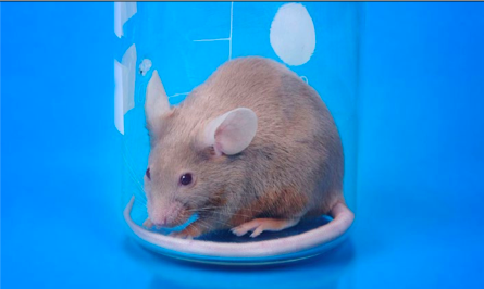
Whilst organisms have their own niche area of research, the most frequently used is the humble mouse. Its prominence is attested by the fact that it was only the second mammalian species after humans to have its genome sequenced, demonstrating that both species share 99% of their genes. [1] Reasons for the popularity of mice as a choice include that mice share many anatomical and physiological similarities with humans. Other advantages include that they are small, hardy, cheap to maintain and easy to breed with a short lifespan (approximately three years), [2] allowing experiments to gather results more quickly. Common human diseases such as diabetes, heart disease, and cancer affect mice, [3] hence complex pathophysiological mechanisms such as angiogenesis and metastasis can be readily demonstrated. [2] Above all, the extraordinary ease with which mice are manipulated has resulted in the widespread availability of inbred, mutant, knockout, transgenic or chimeric mice for almost every purpose conceivable. [3] By blocking or stimulating the overexpression of specific genes, their role in developmental biology and disease can be identified and even demonstrated in specific organs. [4]

Humanised mice are another step closer in representation of what happens in the human body, thereby increasing the clinical value of knowledge gained from experiments. Humanised mice contain either human genes or tissue allowing the investigation of human mechanisms whilst maintaining an in vivo context within the animal. Such approaches are also available in other organisms such as rats, but are often adapted from initial advances in mice, and hardly mirror the ease and diversity with which humanised mice are produced.

Aside from the mouse, invertebrates such as the Drosophila vinegar fly [5] and Caenorhabditis elegans worm [6] are also widely used in research of genetics or developmental biology studies. They are particularly easy to maintain and breed and therefore large stocks can be kept. Furthermore, there are fewer ethical dilemmas and invertebrates have a genome simple enough to be investigated in its entirety without being cost-prohibitive or requiring an exhaustive set of experiments. Their anatomies are also distinct and simple, allowing developmental changes to be readily visualised.

Another alternative is the Zebrafish, which shares many of the advantages offered by Drosophila and C. elegans. Additionally, it offers greater scope for investigating more complex diseases like spinal cord injury and cancer, and possesses advanced anatomical structures as a vertebrate. [7] Given the inherent capacity of the Zebrafish for cardiac regeneration, it is also of interest in regenerative medicine as we seek to harness this mechanism for human therapy. [8]

Large animals tend to be prohibitively expensive, time-consuming to manage and difficult to manipulate for use in basic science research. Instead, they have earned their place in preclinical trials. Their relatively large size and physiological similarity to humans provides the opportunity to perform surgical procedures and other interventions on a scale similar to that used clinically. Disease models created in sheep or swine are representative of the complex biological interactions that are present in highly evolved mammals; hence may be suitable for vaccine discovery. [9] Furthermore, transgenic manipulation is now possible in non-human primates, presenting an opportunity to develop humanised models. [10] Despite this, there are obvious limitations confining their use to specialised settings. Large animals need more space, are difficult to transport, require expert veterinary care, and their advanced psychosocial awareness raises ethical concerns. [9]

The clinical context of animal experimentation

A major issue directly relevant to clinicians is the predictive value of animal models. Put simply, how much of research using animals is actually clinically relevant? Although most medical therapies in use today were initially developed using animal models, it is also recognised that many animal experiments fail to reproduce their findings when translated into clinical trials. [11] The reasons for this are numerous, and require careful analysis.

The most obvious is that despite some similarities, animals are still animals and humans are humans. Genetic similarities between species as seemingly disparate as humans and mice may lead to assumptions of conserved function between humans and other animal species that are not necessarily correct. Whilst comparing genomes can indicate similarities between two species such studies are unable to capture differences in expression or function of a gene across species that may occur at a molecular level. [12]

The effectiveness and clinical relevance of experimental animal trials is further complicated by epigenetics. Epigenetics is the modification of genetic expression due to environmental or other cues without actual change in DNA sequence. [13] These changes are now considered just as central to the pathogenesis of cancer and other conditions as genetic mutations.

It is also important to consider the multi-factorial nature of human diseases. Temporal patterns such as asymptomatic or latent phases of disease can further complicate matters. Patients have co-morbidities, risk factors, and family history, all of which contribute to disease in a way that we may still not completely understand. With such complexity, animal models do not encapsulate the overall pathophysiology of human disease. Animals may be too young, too healthy, or too streamlined in sex or genetics. [14] To obtain animals with specific traits, they are often inbred such that two animals in the same experiment will have identical genetic make-up – like twins, hardly representative of the diversity present in nature. Understandably, it can be an extraordinary challenge to incorporate all these dimensions into one study. This is especially so when the very principles of scientific method dictate that variables except for the one under experimentation should be minimised as much as possible.

A second area of concern is the sub-optimal rigour and research design of animal experiments. Scientists who conduct animal experiments and clinicians who conduct clinical trials often have different goals and perspectives. Due to ethical and cost concerns, the sample size of animal experiments is often kept to a minimum, and studies are prolonged no more than necessary, often with arbitrarily determined end-points. [14] Randomisation, concealed allocation, and blinded outcome of assessment are poorly enforced, leading to concerns of experimental bias. [11] Additionally, scientific experiments are rarely repeated due to an emphasis on originality, whereas clinical trials are often repeated (sometimes as multi-centre trials) in order to assess reproducibility of results. Furthermore, clinical trials are more likely to be published regardless of the nature of results; in contrast, scientific experiments with negative findings or low statistical significance often fail to be reported. These gaps highlight the fact that preclinical trials should be expected to adhere to the same standards and principles of clinical trials in order to improve the translatability of results between the two settings.

Although deficiencies in research conduct is a concern, the fundamental issue that remains is that even the best-designed preclinical study cannot overcome the inherent differences that exist between animal models and ‘real’ human patients. However, it is reassuring to know that we are becoming better at manipulating animal models and enhancing their compatibility with their human counter-parts. As such, this drive towards increasingly sophisticated animal models will provide more detailed and clinically relevant answers. Additionally, with the recognition that a single animal model is inadequate on its own, experiments may be repeated in multiple models. Each model will provide a different perspective and lead to the formation of a more comprehensive and balanced conclusion. A suggested structure is to start initial proof-of-principle experiments in small, relatively inexpensive and easily manipulated animals, and then scale up to larger animal models.

‘Human’ experimental models – the revolution of stem cells

Given the intrinsic differences between animals and humans, it is crucial to develop experimental systems that simulate human biology as much as possible. Stem cells are ‘master cells’ with the potential to differentiate into more mature cells, and are involved in the development and maintenance of organs through all stages of life from an embryo (embryonic stem cells) to adult (tissue-specific stem cells). [15] With the discovery of human embryonic stem cells [16] and other tissue-specific stem cells [17] it is now possible to appreciate the developmental biology of human tissues and organs in the laboratory. Stem cells may be studied under various controlled conditions in a culture dish, or even implanted into an animal to recapitulate in vivo conditions. Furthermore, stem cell transplantation has been used in animal models of disease to replace lost or damaged tissue. These methods are now commencing high-profile clinical trials with both embryonic stem cells [18] and tissue-specific stem cells. [19] Although stem cells hold great potential, translating this into the clinical environment has been hindered by several obstacles. Chiefly, tissue- specific stem cells are rare and difficult to isolate, while embryonic stem cells can only be created by destroying an embryo. In order to generate personalised embryonic stem cells for cell therapy or disease modelling, they need to be created via ‘therapeutic cloning.’ The considerable ethical quandary associated with this resulted in a field mired in controversy and political debate. This led to research coming almost to a standstill. Fortunately, stem cell research was rejuvenated in 2007 with the revolutionary discovery of induced pluripotent stem (iPS) cells – a discovery notable enough to be awarded the 2012 Nobel Prize in Physiology/Medicine.

Induced pluripotent stem (iPS) cells are created by reprograming mature cells (such as skin fibroblasts) back into a pluripotent ‘stem cell’ state, which can then re-differentiate into cells of any of the three germ layers irrespective of what its original lineage was. [20] Cells from patients with various diseases can be re-programmed into iPS cells, examined and compared to cells from healthy individuals to understand disease mechanisms and identify therapeutic opportunities. Rather than using models created in animals, this approach represents a ‘complete’ model where all genes contributing to a specific disease are present. Crucially, this enables the previously inconceivable notion of deriving patient-specific ‘disease in a dish’ models, which could be used to test therapeutic response. [21] It also provides unprecedented insight into conditions such as those affecting the heart [22] or brain, [23] which have been difficult to study due to limitations accessing tissue specimens and conducting experiments in live patients.

However, if a model system rests purely on stem cells alone this would relegate the approach to in vitro analysis without the whole organism outlook that animal experiments afford us. Accordingly, by combining this with rapidly evolving cell transplantation techniques it is possible to derive stem-cell based animal models. Although this field is flourishing at an exponential rate it is still in its infancy. It remains to be seen how the actual translation of iPS technology will fit into the pharmacological industry, and whether personalised drug screening assays will become adopted clinically.

Conclusion

Experimental models provide us with insight into human biology in ways that are more detailed and innovative than ever before, with a dazzling array of choices now available. Although the limitations of animal models can be sobering, they remain highly relevant in biomedical research. Their contribution to clinical knowledge can be strengthened by refining models to mimic human biology as closely as possible, and by modifying research methods to include protocols similar to that used in clinical trials. Additionally, the emergence of stem cells has shifted current paradigms by introducing patient-specific models of human development and disease. However, it should not be seen as rendering animal models obsolete, but rather a complementary methodology that should improve the predictive power of preclinical experiments as a whole.

Understanding and awareness of these advances is imperative in becoming an effective researcher. By applying these models and maximising their potential, medical students, clinicians and scientists alike will enter a new frontier of scientific discovery.

Conflict of interest

None declared.

Correspondence

k.yap@amsj.org


Categories
Book Reviews Articles

Harrison’s: Friend or Foe?

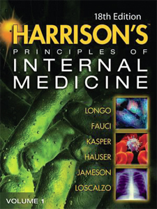
Longo DL, Harrison TR. Harrison’s Principles of Internal Medicine, Eighteenth Edition. London: McGraw-Hill; 2012.

RRP: $199

So a review of this text has been done before, not of Harrison’s Principles of Internal Medicine (Harrison’s) in isolation but a comparison to William Osler’s The Principles and Practice of Medicine. [1] The latest edition of Harrison’s has been available since July 2011, and as an avid user of the online version of Harrison’s (via AccessMedicine™ through the University’s library website). The book is found in two tomes, a whopping 4012 pages in total. I have a thing for being able to physically hold a book and read it, hence not relying on the online edition, which has been previously compared to the text version, as my internet connection is very erratic and the University has a concurrent users policy. [2]

Alas it was a decision that I do regret (to some extent) as I have since found myself referring to Harrison’s to find an answer to a problem, whether it be electronically via the DVD given with the book or via fl icking through the book itself, and neglecting some other general medicine or specialised texts that I own. This speaks volumes about Harrison’s comprehensive nature, but also about my enjoyment of the text.

So what do I like about the book? It is detailed, this may speak more about myself than the text but I think that many medical students appreciate this level of detail, if only for interest rather than what is actually required. I mean, do you know of any other books with 395 chapters and another 51 chapters available electronically? I love the detailed explanations of concepts such as “Insulin biosynthesis, secretion and action”, which would normally be found in a more specialised text such as Lehninger’s Principles of Biochemistry™, and pathophysiology of common diseases such as asthma, COPD and myocardial infarction. [3]

The “yellow sections” in the chapters are a great reference for medical students and physicians alike, these are the sections on treatment of certain conditions. The diagrams are great, as are flowcharts, which explain key concepts such as development of a certain condition (for example, ischaemic stroke) or treatment or diagnostic algorithms, such as tuberculosis or HIV/AIDS. The layout of the parts, sections and chapters of the text are very logical and (if you were keen enough) could be read in order for example:

“Part 10: Disorders of the cardiovascular system, Section 1: Introduction to cardiovascular disorders, Chapter 224: Basic Biology of the Cardiovascular system, Chapter 225: Epidemiology of Cardiovascular disease … Section 2: Diagnosis of Cardiovascular disorders, Chapter 227: Physical examination of the cardiovascular system, Chapter 228: Electrocardiography … Section 3: Disorders of rhythm … Section 4: Disorders of the heart … Section 5: Vascular disease”

It is easy to see how logically the book is organised, starting from the basics of the given system or group of conditions then working through epidemiology, diagnosis and then fi nally about the conditions themselves; and given that Part 10 of the book as a whole spans pages 1797 – 2082 (yes, 285 pages) you can gather an appreciation for the detail of the text. Another great feature is the “further readings” given at the end of each chapter citing original and review publications from peer reviewed journals so (if interested) you can read some more about the topic you are interested in.

What don’t I like about the book? Having two volumes can sometimes be a little tedious when you pick up one and then find that the topic you want is in the other (although you have to remember page numbers this way, it is still preferable to having one enormous tome with a tiny typeface). The organisation of the text is a double-edged sword as it can get frustrating as when searching for a condition such as polycystic ovarian syndrome (PCOS) this will bring up entries in sections such as: menstrual disorders, biology of obesity, amenorrhoea, metabolic syndrome, hirsutism and virilisation and diabetes mellitus; yet there is no definitive section on PCOS itself as there is for a condition such as phaeochromocytoma. Sometimes you open a page, and the amount of text overwhelms you and there are no figures to break it up, which can be quite intimidating for a medical student to find one specific passage or sentence. This isn’t too large a problem in my opinion, but I have known students to be put off by books of such a nature.

References

[1] Hogan DB. Did Osler suff er from “paranoia antitherapeuticum baltimorensis”? A comparative content analysis of The Principles and Practice of Medicine and Harrison’s Principles of Internal Medicine, 11th edition. CMAJ. 1999 Oct;161(7):842-5.

[2] DeZee KJ, Durning S, Denton GD. effect of electronic versus print format and different reading resources on knowledge acquisition in the third-year medicine clerkship. Teach Learn Med. 2005;17(4):349-54.

[3] Powers AC. “Insulin Biosynthesis, Secretion and Action” from Harrison’s Principles of Internal Medicine. 18 ed. Longo DF, A; Kasper, D; Hauser, S; Jameson, J L; Loscalzo, J, editor. Columbus: McGraw-Hill Medical; 2011.

Categories
Articles Editorials

The clinician-scientist: Uniquely poised to integrate science and medicine

Introduction

Growing in the world of academic medicine is a new generation of doctors known as “clinician-scientists”. Trained in both science and medicine, with post-graduate research qualifications in addition to their medical degree, they serve as an essential bridge between the laboratory and clinic.

The development of sophisticated experimental approaches has created opportunities to investigate clinical questions from a basic science perspective, often at a cellular and molecular level previously impossible. With new and detailed understanding of disease mechanisms, we are rapidly accelerating the discovery of new preventative measures, diagnostic tools, and importantly, novel therapeutic approaches. In these emerging avenues there is not just a need for collaboration between scientists and clinicians, but a need for individuals who are fluent in both science and medicine – hence, the advent of clinician-scientists. The terms “translational research” or “translational medicine” are often associated with clinician-scientists, alluding to the notion that these people facilitate the two-way process of translating scientific findings into clinical applications (bench-to-bedside), and provide clinical data and specimens back to the laboratory to investigate underlying disease processes (bedside-to-bench).

From a student’s perspective however, these concepts can be confusing and finding their way through the breadth and categories of research conducted in academic institutions and hospitals may prove daunting. A discussion of the clinician-scientist niche and some of the challenges and opportunities faced may prove helpful.

Defining the clinician-scientist

Most clinicians at an academic hospital are engaged in research to some extent, but this tends to be mainly clinically-oriented, with patient care, treatment outcomes, and population health being broad areas commonly involved. Their day-to-day job is mostly defined by their clinical duties, often with some teaching responsibilities involved. Clinician-scientists, by contrast, dedicate a significant proportion of their time to research, typically spending ≥50% protected time in order to be remain academically competitive. [1] Whilst still loosely defined, in a purist sense this is a clinician who is involved in research at an organ, tissue, cellular, or molecular level, as opposed to focussing solely on whole patients as a clinical subject. Such research may not always have clinical findings that are directly relevant to everyday medical practice but the difference from a pure basic scientist is that the science has been approached with clinical relevance in mind. Interestingly, on the other hand, science itself has become inter-disciplinary and is recognising the importance of clinical relevance and translation with new ventures such as the Stanford University PhD in Stem Cell Biology where graduate science students interested in involvement with translational research in regenerative medicine undertake rotations shadowing clinicians in order to develop a clinical perspective to their research. [2] These developments indicate that not only are the frontiers between science and medicine becoming blurred, but that translational research is the exciting intersection where clinician-scientists, as well as scientists well-attuned to clinical practice, are uniquely poised to thrive.

The clinician-scientist niche

Clinician-scientists possess a distinctive set of skills, being trained as a clinician to apply scientific knowledge to patient care, and trained as a scientist with an enquiring mind designed to test hypotheses. Understanding the clinical relevance of observations in science and the ability to translate this back into clinical practice is truly the domain of the clinician-scientist, and uniquely so.

The pursuit of additional post-graduate research qualification such as a Masters or PhD has traditionally been the main pathway to becoming a clinician-scientist in Australia, unlike in the United States where combined MD-PhD programs have been well established in the past. However, the recent development of similar combined MBBS-PhD and MD-PhD programs in Australia is likely be instrumental in building a body of clinician-scientists that have been moulded specifically for this task. [3] Skills developed in scientific training essential for success in research include literature appraisal, manuscript and grant writing, and mastery of laboratory techniques, all of which are life-long skills honed over time, and which are rarely acquired in medical school.

It goes without saying that clinician-scientists are expected to be experts in both medicine and science. Anything subpar of clinical competence would pose a threat to patient safety and cannot be compromised. On the other hand without a solid commitment in research with the appropriate output in terms of publications, conference attendance, and grant proposals, a career in research will not take off since a track record is something that needs to be built on constantly. Given that clinical training itself takes a good number of years before being able to practice as an independent clinician it is little wonder that many are unwilling to tackle both clinical and scientific careers at once. Again, this lends further credence to the MD-PhD path where scientific training would have already been completed by the end of the program, although this itself has its drawbacks, since the science gained can become neglected in the last clinical years and will need to be polished again upon completion. [4]

But where lie challenges also lie opportunities: for the determined few, funding statistics indicate that the rigorous training is entirely worthwhile. Clinician-scientists have been found to consistently perform better in national funding programs such as the National Institutes of Health Research Project Grants (United States) than their pure clinician (MD only) and basic science (PhD only) counterparts. [5] Although the pool of clinician-scientists in Australia is significantly smaller than that of the United States and data on funding trends are less widely discussed in literature, it is generally acknowledged that clinician-scientists also do well in obtaining NHMRC funding. This may be due partly to the fact that clinician-scientists are afforded more flexibility in labelling their projects as “basic science” or “clinical”, and therefore have access to funds for both basic science and clinical projects, whereas pure clinicians and scientists are generally limited to their own funding areas.

When describing the clinician-scientist niche, an aspect of research “translation” that is often neglected is the importance of the delivery of research-based medicine into actual practice. The classic bench-to-bedside process refers to the invention of a new drug, device, or diagnostic tool where the hope is that it will undergo clinical evaluation in a controlled setting with a specific patient cohort. But bringing a discovery into the market is simply the beginning, and to bring this to the general public a much more concerted effort is required involving collaboration between public health experts, policy makers, and clinicians amongst others. So drawn-out and complex is the process that it is well acknowledged that this area of “translational” research often fails, with many potentially important discoveries unable to make changes to everyday medical practice.

[6] However, clinician-scientists are well suited to play an active role in negotiating the many hurdles in this endeavour by facilitating communication between the various experts involved, whilst providing a first-hand inventor as well as treating clinician’s perspective that is not only unique but critical in ensuring that an invention is appropriately implemented and evaluated. In the Australian context, the National Health and Medical Research Council (NHMRC) has recognised this gap in research translation and the Centres for Research Excellence and Translating Research Into Practice (TRIP) Fellowships are specific measures aimed to address this issue. [7]

Wearing two hats: double the challenges?

A commonly quoted recommended research:non-research ratio for workload is 75:25, with the majority of time devoted to research in order to succeed as a clinician-scientist. [8] In reality this is more likely to be exactly opposite the case, where a 75:25 ratio in favour of clinical work becomes the norm instead. [9] This may be particularly so in the early years after graduation when specialist training is being undertaken, despite the fact that this is also the time when a solid research foundation needs to be built in order to establish a clinician-scientist’s academic presence. As pressing as clinical demands may be, it is widely recognised that a research career cannot flourish without negotiating some protected time from clinical duties with the hospital department.

The biggest challenge for clinician-scientists is therefore time management. In addition to patient care, clinical training, and teaching responsibilities, clinician-scientists are expected to undertake labwork, keep abreast of advances in both scientific and medical literature, and engage in professional development and conferences on both fronts. They must maintain manuscript preparation and grant proposals, complete administrative duties, and often lead research teams. To realistically keep up with these demands of juggling a dual career, the ability to delegate and seek cooperation from scientist and clinician colleagues is critical. The lack of a supportive environment and a suitable mentor who can share their experiences and show the way can present an impossible struggle to the time-constrained clinician-scientist.

On the clinical front, to manage their workload clinician-scientists may tightly focus their interests to subspecialised areas to maintain an adequate caseload and expertise without stretching oneself too thin. This depends however on working in an environment where the volume and diversity of patients permits such subspecialisation, with appropriate facilitation by supervisors such as Department Heads. Unfortunately these conditions tend to be found only in major tertiary hospitals, relegating clinician-scientists to these settings.

Additionally, a research career is often less financially rewarding than clinical work particularly when private practice may need to be sacrificed in order to undertake lab work. This can pose a significant barrier particularly because the number of years required to gain appropriate training results in clinician-scientists being likely to be older than their scientist and clinician counterparts and may therefore have family commitments, and have often also accumulated student debts that need to be repaid. [10] Some solutions to this may be the Practitioner and Career Development Fellowships offered by the NHMRC aimed at clinicians involved in research, [11] as well as hospital and philanthropic organisation funding specifically for buying time out from clinical practice for research.


Opportunities for the clinician-scientist


For any researcher, securing funding is a lifeline in continuing their work and burnishing a track record, and it is here where clinician-scientists can be creative in sourcing their benefactors. Philanthropic organisations often affiliated with a disease or clinical cause, specialist training colleges like the Royal Australasian College of Surgeons, hospital based foundations, pharmaceutical companies, and fundraising from patient advocates are all important and significant funding avenues that clinician-scientsts may find more accessible than pure scientists. [12] These grants often allow pilot projects to be undertaken in order to generate sufficient amount of preliminary data to become competitive for major research funding such as from the NHMRC. Additionally, a number of these organisations offer clinician-scientist fellowships similar to the NHMRC.

Apart from funding success, it has also been found that many clinician-scientists opt to apply for and are successful in obtaining university academic positions. [12,13] Such engagement in academia provides synergy for research efforts by opening up institutional resources often more diverse than hospital settings, prospects for networking with likeminded professionals and mentors.

Additionally, the scope translational research itself is widening. An increasing number of academic hospitals are dedicating departments to translational research, with clinician-scientists often taking the lead. The need to prioritise translational research has been further underlined by the Chief-Scientist of Australia’s recent speech calling for increase in research funding for this area. [14] Whilst these are positive developments, further input from clinician-scientists themselves is required to shape policy changes and design steps to increase their numbers.


Moving forward


An apt saying may be, “Clinicians know all of the problems, but none of the solutions; scientists know all of the solutions, but none of the problems”. [15] This is where clinicianscientists represent a unique breed suited to fulfil this vacant niche, and are absolutely necessary in forging the next success stories of medicine. Despite the complexities of a dual career, the rewards and satisfaction in pursuing this path are evident and meaningful, and can lead to tangible health outcomes in patients. Although it is important to maintain a realistic notion that being a clinician-scientist is by no means an easy feat, it is equally important to take hope that the best of both worlds can be experienced. These perspectives are increasingly acknowledged in the form of progresses being made in the right direction to encourage clinician-scientists. In light of this, perhaps it is well worth noting that there may never be a better time than now to venture into, and indeed take charge in riding this next wave of medical evolution.

References

[1] Archer SL. The making of a physician-scientist–the process has a pattern: lessons from the lives of Nobel laureates in medicine and physiology. Eur Heart J. 2007 Feb;28(4):510-2

[2] Stanford University School of Medicine, Institute for Stem Cell Biology and Regenerative Medicine. PhD Program: Curriculum Overview. [updated February 17th 2009; cited March 8th 2012]; Available from: http://stemcell.stanford.edu/education/phd/curriculum.html

[3] Power BD, White AJ, Sefton AJ. Research within a medical degree: the combined MB BS-PhD program at the University of Sydney. Med J Aust. 2003 Dec 1-15;179(11-12):614-6

[4] Marban E, Braunwald E. Training the clinician investigator. Circ Res. 2008 Oct 10;103(8):771-2

[5] Dickler HB, Fang D, Heinig SJ, Johnson E, Korn D. New physician-investigators receiving National Institutes of Health research project grants: a historical perspective on the “endangered species”. JAMA. 2007 Jun 13;297(22):2496-501

[6] Woolf SH. The meaning of translational research and why it matters. JAMA. 2008 Jan 9;299(2):211-3

[7] McCallum J, Forster R. From the NHMRC: Research translation network targets the evidence-practice lag. Med J Aust. 2011;195(5):252

[8] Tai IT. Developing a clinician-scientist career. Clin Invest Med. 2008;31(5):E300-1

[9] Bosse D, Milger K, Morty RE. Clinician-scientist trainee: a German perspective. Clin Invest Med. 2011;34(6):E324

[10] Lander B, Hanley GE, Atkinson-Grosjean J. Clinician-scientists in Canada: barriers to career entry and progress. PLoS One. 2010;5(10)

[11] National Health and Medical Research Council. Fellowship Awards. [Internet] [updated December 21st 2011; cited March 1st 2012]; Available from: http://www.nhmrc.gov.au/grants/apply-funding/fellowship-awards

[12] Hayward CP, Danoff D, Kennedy M, Lee AC, Brzezina S, Bond U. Clinician investigator training in Canada: a review. Clin Invest Med. 2011;34(4):E192

[13] Toouli J. Training surgeon scientists. ANZ J Surg. 2003 Aug;73(8):630-2

[14] Australian Government – Chief Scientist of Australia 2012. Can Australia afford to fund translational research? [updated April 3rd 2012]; Available from: http://www.chiefscientist.gov.au/2012/04/can-australia-afford-to-fund-translational-research/

[15] Hait WN. Translating research into clinical practice: deliberations from the American Association for Cancer Research. Clin Cancer Res. 2005 Jun 15;11(12):4275-7

Categories
Original Research Articles

Onsite and offsite use of computer aided learning in undergraduate radiology education

Aim: Computer-aided learning (CAL) is considered comparable to traditional media for undergraduate radiology teaching. Previous studies have often compared the efficacy of traditional media to onsite CAL use, yet real world usage of CAL is likely to occur in offsite settings. This study aims to compare usage and learning outcomes of a chest radiology CAL in onsite and offsite settings. Methods: Participants were fourth year medical students (n=52) at the National University of Singapore (NUS) undertaking one week radiology rotations. Students were randomly allocated to complete a web-based chest radiology CAL onsite, or offsite at a time and place of choice. Pre- and post-tests were taken to measure knowledge gain, and a questionnaire was used to explore student usage and preferences.

Results: The onsite CAL group demonstrated significant knowledge gain (+15.8%, p<0.05) whilst the offsite group did not (+5.8%, p>0.05). However, the difference between the groups was not statistically significant (p=0.069). Total time spent and completion of the program was similar between the two groups. Yet, questionnaire results showed that the offsite group multitasked more and appeared to have poorer concentration. A majority of students from both groups preferred the convenience of offsite CAL use over onsite CAL use.

Conclusion: A significant difference between the test groups was not observed, although there was a trend toward onsite CAL use being more effective. In planning CAL teaching, particularly for offsite use, educators need to provide sufficient support and integration for an optimal outcome.

Introduction

Chest radiology is important for acute and emergency management, and is therefore an essential learning component of undergraduate radiology teaching. [1] However, studies show that chest radiology competency amongst graduating medical students is poor. [2,3] Poor competency is attributed to lack of formal teaching of radiology in the curriculum. [2,3] Worldwide, radiology teaching is compromised by limited formal teaching in a hectic curriculum, and competing demands on radiologists. [4,5]

Computer aided learning (CAL) has been advocated as a potential tool to alleviate some of the limitations in radiology teaching. [6] CAL is time and cost effective for educators, [7] and especially useful in an image rich specialty such as radiology. To evaluate the effectiveness of CAL for transferring knowledge gain, previous studies have undertaken media comparisons between CAL and traditional learning, such as lectures or tutorials. Individual studies in radiology and non-radiology medical education [8,9] demonstrate that overall, knowledge gain with CAL is comparable to …

Categories
Review Articles

The significance of aphasia in neurological cancers

Abstract

Aphasia associated with brain tumours has previously been regarded as essentially equivalent to the aphasia of stroke, and as a deficit unlikely to affect a patient’s prognosis. Recent research challenges such hypotheses. Tumour-related aphasias are commonly anomic aphasias, and hence pathologically distinct from classic post-stroke aphasias. Accordingly, many rules from the world of stroke cannot be readily translated to the management of tumour-related aphasia. Furthermore, aphasia may be an important clinical prognostic parameter in neuro-oncology. Tumour-related aphasia is associated with an increased risk for developing depression, poorer coping and reduced survival time. It is important that health professionals are aware of the unique pathology and prognostic significance of neuro-oncological aphasia, and of strategies available for its relief.

Categories
Feature Articles

A very good iDEA: The inaugural gathering of the student division of Doctors for the Environment Australia

The result of one attendee’s bright iDEA.

In early December 2009, just prior to the much-hyped COP15 round of United Nations climate negotiations in Copenhagen, 40 medical students, representing six states and eleven medical schools, descended upon Melbourne for iDEA, the inaugural gathering for the student division of Doctors for the Environment (DEA). Attendees were encouraged to be mindful of their carbon footprints whilst travelling to the conference, with many students opting for train or coach rather than air travel. Most impressively, three Tasmanians cycled for three days from Hobart to Melbourne University (with the assistance of the Bass Strait ferry).

Education and networking were the focus of this three day gathering at Newman College within the University of Melbourne, where a plethora of distinguished speakers presented talks and interactive workshops to enlighten the receptive minds in attendance: academics, environmental activists, clinicians and all combinations of the three.

All present agreed that it was long overdue that medical students gathered to discuss environmental issues relevant to health; issues that for various reasons have been sidelined by the medical fraternity. These issues often traverse traditional subject boundaries, implying a perceived or real lack of academic expertise. Additionally, the lack of confidence in using one’s ‘authority’ as a medical professional plays a part. Climate change, for instance, is often seen as a political or economic concern rather than a threat to health. Being too busy, self-preservation, fear over allegations of hypocrisy, ignorance, inertia and ‘donor fatigue’ all contribute to the reluctance of doctors to speak up.

According to Costello et al., climate change “is the biggest global health threat of the 21st century” and the repercussions to health will be global in reach, but with a disproportionately large impact falling on the developing world. [1] Matthew Wright, co-founder of Beyond Zero Emissions, a Melbourne-based organisation promoting the rapid transition to a zero carbon future, raised the interesting point that planning for a zero-carbon future is different to planning for a low emissions future, which, in turn, is different to planning for a doubtful emission reduction trading scheme in which concessions are made to big polluters. Although it seems paradoxical, government inaction in the short term could thus be preferable to legislating a hurried, binding scheme, that is in fact ineffectual in preventing an unsafe average global warming of two or more degrees.

Richard Di Natale, a former GP and Public Health physician, provided insight into how one might make the transition from clinician to environmental activist and politician. His non-linear career trajectory has seen him transition through positions in primary care, HIV programme development, Government Health Department bureaucracy and community-building. Most recently, he is persuading Victorian voters to give him the job of a Greens Senator at the next Federal election…

Categories
Review Articles

What do medical students think about pharmaceutical promotion?

Abstract

Aim: The aim of this review was to produce an overview of surveys of medical students’ exposure to and attitudes towards pharmaceutical promotion. Methods: PubMed was searched for studies featuring surveys of medical students regarding their interactions with pharmaceutical promotion and tabulated the findings for survey questions relating to the main themes. Results: Students have significant exposure to promotion, and they generally view receiving gifts as acceptable, but do regard some gifts as more appropriate than others. Most students think pharmaceutical sales representative (PSR) presentations are biased but still of educational value and should not be banned. Most students do not believe promotion will affect their prescribing behaviours. A large majority of students want more education in their curricula on how to interact with PSRs. Conclusions: Many medical students think that pharmaceutical promotion is biased and feel underprepared for interactions with the pharmaceutical industry. Despite this, they accept exposure to pharmaceutical promotion believing that it will not influence them. There is scope for improved education in medical schools about this issue.