Categories
Guest Articles

For Euthanasia: Murder versus mercy

Under existing law, if a doctor intentionally and compassionately hastens a patient’s death, upon the patient’s request to end their suffering, that doctor can be prosecuted with the most serious of crimes – murder.

Is this a just law? If you answer ‘No . . . the doctor should not be charged with murder’, then you favor law reform for voluntary euthanasia (VE). If you say ‘Yes . . . the doctor should be prosecuted for murder’, then you are against VE reform.

Surveys of Australians over the past few decades consistently show that around 80% believe a person with terminal illness should have a legal option for VE to end their suffering. [1] About 50% of doctors favor law reform. [2] Curiously, it seems palliative clinicians and parliamentarians have the lowest level of support.

I came into palliative medicine with no fixed position on VE, but as I witnessed the suffering of patients, and listened to their wishes, some pleaded with me to hasten their demise. I progressively formed a view in favor of VE reform.

In this short article, I will examine some of the major myths about VE. I will allude to published evidence, including from jurisdictions where either VE or physician assisted suicide (PAS) has been legalized (The Netherlands, Belgium, Luxembourg, Switzerland, and USA – Oregon, Montana and Washington).

Myth: Palliative care relieves suffering so there is no need for VE18

The problem is that it is impossible to relieve all suffering. Dying people have varied and sometimes intense suffering, including physical, emotional, and existential suffering. Every survey of hospice patients shows they experience multiple concurrent symptoms. Severe refractory symptoms, including suffocation, pain, nausea and confusion, requiring palliation with deep sedation, have been reported in up to 50% of palliative care patients. [3]

Also, surveys show 5 to 10% of patients with advanced cancer request a hastened demise, and this proportion is actually higher in patients who receive hospice care. [4,5]

In the VE debate, many palliative specialists have difficulty hearing and representing the wishes of their patients who want VE, and difficulty appraising the relevant literature. Does some ideology (perhaps medical or religious) impede their ability? Do they fear being personally involved in VE? Why do they view VE as antagonistic to palliative care?

Myth: Legalizing VE undermines palliative care development

In fact, the opposite occurs – when VE legislation is introduced, palliative care is boosted. For example, in the Northern Territory, as a consequence of debate about The Rights of the Terminally Ill Act, the world’s first VE legislation, the NT palliative care service gained the highest per capita funding in Australia. In Oregon, where PAS was enacted in 1997, palliative referrals increased, markers of end-of-life care became among the best in the USA, and over 90% of those who accessed PAS also had hospice care. [6,7]

When VE reform is being considered, the importance of palliative care becomes obvious to parliamentarians and health care administrators, so its development is naturally enhanced rather than undermined.

Myth: Allowing VE creates a ‘slippery slope’

This argument suggests doctors will develop a ‘lust for killing’, that ‘a culture of death’ will grow, and vulnerable people will increasingly be pressured to die, or be killed against their wishes (a bleak view of humanity!). Data from more and more jurisdictions, however, indicate the ‘slippery slope’ is a myth.  For example, in the Netherlands, 1.7% of deaths involved VE in 1990, and in 2005 it was the same (1.7%). [8] In Oregon, where 0.2% of all deaths involve PAS, it is educated rather than vulnerable persons who access PAS. [6,7]

Interestingly, surveys in countries that have yet to sanction VE or PAS have higher rates of (covert) voluntary and non-voluntary euthanasia. For example, a survey of Australian medical practice revealed 3.5% of deaths involved ‘ending life without explicit request’, whereas the comparable figure in The Netherlands was 0.7%. [9]

Also, a survey of Australian surgeons revealed one third had given medications with the intention of causing death, often without request (if the criminal law was thoroughly enacted, there would be a lot of Australian surgeons in prison!). [10]

This raises the possibility that visibility (through reform) may be the best way to protect vulnerable patients.

Myth: Negative effects on the doctor-patient relationship

It is the role of the doctor (conferred by society) to make life-and-death decisions. It is routine for doctors to withdraw and withhold life-prolonging treatments, and to administer medications to relieve suffering, even if death is hastened. Similarly, people want doctors to assist them with VE and PAS.

Doctors in Oregon were more likely to receive an explicit request for assisted suicide if they found caring for a dying patient ‘intellectually satisfying’, if they sought to improve their knowledge of pain control in the terminally ill, and if they were willing to prescribe a lethal medication. [11] Those who opposed PAS were twice as likely to report patients becoming upset, or leaving their practice, as a result of their position compared with physicians who supported PAS. [11]

We should aim to satisfy the wishes and interests of every patient, and to do our best for each individual that seeks medical help. I think this is why some doctors flout the criminal law, at great risk to themselves, to covertly provide VE for patients in unbearable suffering who plead for such help.

Myth: VE is a form of killing that is unethical

Opponents portray VE as a form of immoral killing, yet there are differences between murder and VE, just as there are differences between rape and making love. A valid moral appraisal must take account of the wishes of the subject, the motivation of the act, and its overall context.

Personal liberty underpins VE – the ability of an individual to make an autonomous choice about the end of their life. VE also requires an act of conscience by the doctor, whose motivation should be compassion and mercy for the person who is suffering and requests help to die.

Conclusion

The overwhelming majority of people want to have a choice about ending their life, should they be suffering with terminal illness. However, the proportion of people with terminal illness who actually want to end their lives with VE or PAS is quite small. Palliative specialists cannot eliminate all the harrowing indignity and disintegration of dying, and should acknowledge the wishes of patients for a hastened demise. Sanctioning VE will promote palliative care; it does not create a ‘slippery slope’, nor undermine the doctor-patient relationship. It is misguided paternalism that denies patient choice, a lack of mercy that mandates suffering, and an unjust law that puts doctors at risk of serious prosecution.

References

[1] Morgan Gallup Polls in response to the question: ‘If a hopelessly ill patient, in great pain with absolutely no chance of recovering, asks for a lethal dose, so as not to wake again, should a doctor be allowed to give a lethal dose, or not?’

[2] Dying with Dignity NSW. Medical opinion. (2015). [Internet] Available from: http://www.dwdnsw.org.au/medical-opinion/

[3] Cherny N, Portenoy R. Sedation in the management of refractory symptoms: guidelines for evaluation and treatment. J Palliat Care. 1994 Summer;10(2):31-8.

[4] Hunt R, Maddocks I, Roach D, McLeod A.  The incidence of requests for a quicker terminal course.  Palliat Med. 1995;9(2):167-168

[5] Seale C, Addington-Hall J.  Euthanasia: the role of good care. Soc Sci Med  1995; 40(5):581-7

[6] Oregon Government. Death With Dignity Act Annual Report (2012) [Internet] Available from: http://www.healthoregon.org/dwd

[7] Quill, TE. Legal Regulation of Physician-Assisted Death — The Latest Report Cards. N Engl J Med. 2007; 356:1911-1913

[8] van der Heide A, Onwuteaka-Philipsen BD, Rurup ML, Buiting HM, van Delden JJ, Hanssen-de Wolf JE, Janssen AG, Pasman HR, Rietjens JA, Prins CJ,Deerenberg IM, Gevers JK, van der Maas PJ, van der Wal G. End-of-Life Practices in the Netherlands under the Euthanasia Act. N Engl J Med. 2007; 356; 1957-1965.

[9] Kuhse H, Singer P, Baume P, Clark M, Rickard M.  End-of-life decisions in Australian medical practice.  Med J Aust. 1997, 166; 191-196

[10] Douglas CD, Kerridge IH, Rainbird KJ, McPhee JR, Hancock L, Spigelman AD. The intention to hasten death: a survey of attitudes and practices of surgeons in Australia. Med J Aust. 2001;175(10):511-5

[11] Ganzini L, Nelson HD, Lee MA, Kraemer DF, Schmidt TA, Delorit MA. Oregon physicians’ attitudes about and experiences with end-of-life care since passage of the Oregon Death with Dignity Act. JAMA, 2001; 18; 2363-9.

Categories
Guest Articles

Response to “Murder versus mercy”

There is so much misinformation and outdated information surrounding the debate about assisted dying that it is important to try to ascertain what evidence is currently available. Otherwise, myths tend to become ‘reality.’

Relief of suffering

The notion that suffering can be relieved is an attractive one but surprisingly there has been little work undertaken to identify exactly what is meant by the word. Of course, individual suffering is just that – individual. Eric Cassell emphasised the importance of knowing the patient and their values (such as opinions, attitudes, and hopes) in order to try and understand what their suffering is. [1] In Cassell’s view, the nature of the illness and the way a patient responds to it reflects the nature of the patient. It is the striving to understand the intricacies of each individual person that makes palliative care such a rich and rewarding discipline. In a systematic review, Best et al. [2] revealed that suffering “is multidimensional, oscillating, individual and difficult for individuals to express.” They concluded that “opportunities should be provided for patients to express their suffering. The potential for suffering to be transcended needs to be recognized and facilitated by healthcare staff.” Euthanasia is certainly a short-cut to ending that suffering, but as Best and colleagues suggest, many people do indeed transcend that suffering in their last days or weeks. We should not give up on trying to help them do that.  The idea that severe refractory symptoms causing suffering occur in up to 50% of patients is certainly not my experience over 26 years of practice, and indeed the paper quoted is over 20 years old – many advances have been made over two decades.

The ‘slippery slope’

More recent data suggest that the slippery slope is indeed a reality. In the Netherlands in 2013 there has been a 15% increase in reported deaths. [3] Somewhat alarmingly, there is an increase “in situations of beginning dementia (from 42 people in 2012 to 97 in 2013) and psychiatric diagnoses (from 14 people in 2012 to 42 in 2013).” [3] The Dutch Review Committees write that “there is an apparent increasing readiness amongst physicians to comply with requests in general and those in case of dementia and psychiatry in particular. It remains difficult to  find  an  unambiguous  explanation  for  this  increase  in  numbers of reported cases.” It is also suggested now that around one in five patients choosing euthanasia in the Netherlands act under pressure from family members. Professor Theo Boer, one of the supporters of the legislation in that country and a member of a euthanasia Regional Review Committee, who has now recognised the dangers of legalising euthanasia, is also especially concerned about the extension of euthanasia as an option for children – a similar situation to Belgium. Perhaps not surprisingly, assisted dying has also increased in Belgium (by 25% over three years), Washington State (by 17% over 3 years) and Oregon (by 30% in four years). These figures are only for reported assisted dying. It is estimated that under-reporting of euthanasia in the Netherlands represents 20-23% of all euthanasia deaths. A more recent example of the loosening of restrictions on premature ending of life is the development in the Netherlands of a network of traveling euthanizing doctors under the name ‘End of Life Clinic.’ These doctors do not need a relationship with the patients they see. Will this perhaps just become another easy way to reduce the ‘burden’ on a family or on society? The British Medical Association has previously stated that “any moral stance founded on the permissibility of active termination of life in some circumstances may lead to a climate of opinion where euthanasia becomes not just permissible but desirable.” [4]

Doctor-patient relationship

The involvement of doctors in ending a life will necessarily impact on the doctor/patient relationship. This relationship currently is dependent on mutual trust; however, this bond will become increasingly fragile as doctors seek their boundaries in the issue over life and death changing. Assisted suicide offers no second chances. ‘Terminal’ diagnoses are often wrong or inaccurate in their timing. Perhaps this is why doctors’ groups worldwide are persisting in their opposition of law change. Most recently, the British Geriatric Society (BGS) has published a position paper stating that they “do not accept that legalising physician assisted suicide is in the broader interests of society…older people are often strongly influenced by their families and carers – the vast majority, but not all, will have their wellbeing at heart. Even so, many requests to end life…come from the patients’ families and not the older person themselves.” [5] As the BGS suggest, “crossing the boundary between acknowledging that death is inevitable and taking active steps to assist the patient to die changes fundamentally the role of the physician, changes the doctor-patient relationship and changes the role of medicine. Once quality of life becomes the yardstick by which the value of human life is judged, the protection offered to the most vulnerable members of society is weakened”.

References

[1] Cassell EJ. The nature of suffering and the goals of medicine. N Engl J Med 1982; 306(11):639–645.

[2] Best M, Aldridge L, Butow P, Olver I, Webster F. Conceptual analysis of suffering in cancer: a systematic review Psycho-Oncology 2015, DOI: 10.1002/pon.3795

[3] Regional Euthanasia Review Committees Annual Report 2013 [Internet] [cited 2015 July 16]. Available from: https://www.euthanasiecommissie.nl/Images/Annual %20report%202013_tcm52-41743.pdf

[4] The British Medical Association. What is current BMA policy on assisted dying? [Internet]. 2015 [cited 2015 July 16]. Available from: http://bma.org.uk/practical-support- at-work/ethics/bma-policy-assisted-dying

[5] British Geriatrics Society. Physician Assisted Suicide [Internet]. 2015 [cited 2015 July 16]. Available from: http://www.bgs.org.uk/index.php/specialinterest-main/ethicslaw-2/4067- position-assisted-suicide

Categories
Guest Articles

Against Euthanasia: Assisted dying is not part of good medical practice

The issue of assisting or hastening death is not a new phenomenon.19

In the 5th century BC, Hippocrates explicitly stated that new physicians must refrain from such a practice. [1] In 21st century practice the majority of jurisdictions around the world still uphold that principle. However, there is increasing pressure from some groups of the public to “have the right to die”. What lies behind this movement?

Monforte-Royo et al. [2] undertook a systematic review and meta-ethnography from the perspective of patients to try and find out. Their findings are summarised thus: “that the expression of the wish to hasten death…is a response to overwhelming emotional distress and has different meanings, which do not necessarily imply a genuine wish to hasten one’s death.” Other writers have variously described “total pain”, [3] “demoralization syndrome” [4] and “syndrome of end-of-life despair” [5] as being common near the end of life.

The proliferation of hospice and palliative care services worldwide since the late 1960s means that many people now have access to expert help to address these issues, but sadly, not everyone in the medical profession or the public at large feels comfortable dealing with end-of-life issues. Despite the knowledge that every newly qualified doctor will have to deal with upwards of forty people who are dying and their families, in their first year at work as an intern, our medical schools still put little emphasis on this essential element of medical practice.

It is telling that those in the medical profession who are most opposed to assisted dying are those who deal with people who are dying on a daily basis, the palliative medicine physicians. [6] Palliative care is focussed on making the most of each day of life, relieving the burden of troublesome symptoms and addressing psychosocial and spiritual concerns. If practitioners are not properly trained in these areas it is no surprise that some will feel they have no alternative but to acquiesce to a request to assist someone to die. For many, “allowing practitioners to hasten the death of a patient speaks more of abandonment when patients (and their family) need to be drawn together for higher quality of life until death”. [7]

In the majority of requests for hastened death, fear emerges as a major theme: fear of imminent death and fear of the process of dying. [2] Similarly, many patients see euthanasia as a way to end suffering – “as a way out or as a means of relieving loneliness, fear, dependence, a lack of hope and the feeling that life [was] no longer enjoyable”. [2] In addition, many of these patients see euthanasia as a way of reducing the suffering caused to family and carers. If this is the case, is it not the job of the caring professions at least to attempt to reduce or remove this fear and sense of hopelessness?

Those who work with people near the end of life know what is likely to happen in the process of dying: symptoms can be relieved, explanations can be given, suffering can be addressed and not felt to be too hard to deal with.  Rather than rushing to create legislation in an attempt to address the requests for assisted dying, would it not be better to try and understand the meaning patients in the advanced stages of disease attribute to their suffering and its consequences which render them so vulnerable? And then to ensure that every health care practitioner is equipped to deal with such issues?

Any society can be judged by its ability and willingness to care for those who are most sick and vulnerable. People approaching the end of their life are perhaps amongst the most vulnerable. In addition, an individual’s vulnerability to influence and to be made to feel a nuisance or a burden is not unusual. So, how do we assess competence of people who request a hastened death? Reduced mental capacity is common in acutely ill people and yet it has been suggested that clinicians tend not to recognise incapacity. [8] Even in jurisdictions where assisted dying is practiced, psychiatrists have found it difficult to assess whether or not a patient is depressed.

Should the medical profession be involved in the ending of life at all? Many would argue not. It is well established that, amongst those who have been involved with ending life, feelings of emotional discomfort are relatively common. In one study, a proportion of the doctors involved have reported that the emotional burden of having performed euthanasia or assisted suicide had adversely affected their practice. In many ways this is not surprising, as doctors are trained to preserve life, not to end it. [9,10]

Even in countries where the law is clear that an assisted death is permissible, the practical and ethical issues that result from considering and acting upon a request are complex and troubling for most practitioners. [7] The voice of those who would have to do the killing in these circumstances is rarely heard. Perhaps before much further debate into the creation of legislation to support such moves takes place, the role of doctors in this disturbing practice should be made clear.

To accompany people when they are at their most vulnerable and frightened can be hard work, and it is not something that everyone can or will want to do. Each day brings new challenges and opportunities for the patient, their families and their carers. For those who are sick, it is one of the most challenging times of their lives, and yet, paradoxically, it can be one of the most rewarding. The privilege of working with those people and their families is immense. The job of the palliative care specialist and the family medicine doctor is to guide, reassure and comfort not only those who are dying but also those who love and care for them.  It is the job of those who accompany people who are approaching death to help rekindle hope, minimize fear and never to abandon them.

Euthanasia, or assisted dying in any form (including assisted suicide), is therefore antithetical to the purpose and practice of medicine as a whole and to the practice of palliative care in particular.

References

[1] Edelstein L, Temkin O, Temkin CL. Ancient medicine: selected papers of Ludwig Edelstein. Baltimore: Johns Hopkins; 1967

[2] Monforte-Royo C, Villvicencio-Chavez C, Tomas-Sabado J, Mahtani-Chugani V, Balaguer A. What lies behind the wish to hasten death? A systematic review and meta-ethnography from the perspective of patients. Plos One 2012; 7;5 e37117

[3] Saunders C. The last stages of life. Am J Nurs. 1965; 65: 70-75

[4] Clarke DM, Kissane DW. Demoralization: its phenomenology and importance. Aust NZ J Psych. 2002; 36: 733-742

[5] McClain CS, Rosenfeld B, Breitbart W. Effect of spiritual well-being on end-of-life despair in terminally-ill cancer patients. Lancet 2003; 361:1603-1607

[6] Seale C.  End-of-life decisions in the UK involving medical practitioners. Pall Med 2009; 23(3): 198-204

[7] MacLeod RD, Wilson DM, Malpas P. Assisted or hastened death: the healthcare practitioner’s dilemma. Global J Health Sci 2012; doi:10.5539/gjhs.v4n6p

[8] Sessums LL, Zembrzuska H, Jackson JL. Does this patient have medical decision-making capacity? JAMA 2011; 284(19): 2483-2488

[9] Georges JJ, The AM, Onwuteaka-Philipsen BD, van der Wal G. Dealing with requests for euthanasia: a qualitative study investigating the experience of general practitioners. J Med Ethics 2008; 34(3): 150155

[10] Emanuel EJ, Daniels ER, Fairclough ER, Clarridge BR. The practice of euthanasia and physician-assisted suicide in the United States. JAMA 1988; 280(6): 507-513

Categories
Guest Articles

Response to “Assisted dying is not part of good medical practice”

I wish to make several points in response:

Relief of suffering

My colleague claims that with ‘expert’ palliation ‘symptoms can be relieved, explanations can be given and suffering can be addressed and not felt to be too hard to deal with’. This is an idealised view that does not acknowledge the limitations of palliative care, and promotes the myth that suffering in terminal illness can be relieved to the extent there should be no desire nor need for voluntary euthanasia/physician assisted suicide (VE/PAS).

In reality, it is not possible to effectively relieve fatigue and dependency, or eliminate symptoms caused by failing organs, or change many patients’ minds. Evidence indicates that patients receiving specialist palliative care are only marginally better off in terms of symptoms, yet express the wish for a hastened demise more-so than other dying patients. [1]

Patient autonomy

My colleague highlights that patients’ requests for VE/PAS are due to  ‘overwhelming  emotional  distress’,  ‘total  pain’,  ‘hopelessness’, ’demoralisation’, ‘despair’,     ‘fear’,     ‘loneliness’,     ‘vulnerability’, ‘depression’ etc. The argument is that their decision-making capacity is lacking, so there is no need to respect their wishes.

Requests for a hastened demise, however, can be genuine, rational, and in accordance with long-held life values. A person’s autonomy is not invalidated because of their suffering.

It is inconsistent and perplexing when respect for patient autonomy is promoted as a core value of palliative care, including for the withholding and withdrawing of life-prolonging treatments, but not when it applies to VE/PAS.

Effect on clinicians

My colleague suggests that clinicians who become involved VE/PAS are ill-equipped and uneducated in palliative care, and they are adversely affected by ending life.

In Oregon, however, doctors who are actively interested in palliation at the end of life are more likely to be involved in PAS. [2]

Accompanying terminally ill patients with compassion, with respect for their autonomy (the opposite of abandonment), can lead some doctors to be involved in PAS/VE. To borrow the words of my colleague, this “can be hard work, and it is not something that everyone can or will want to do . . . it is one of the most challenging times  . . . yet paradoxically it can be one of the most rewarding.”

Voluntary euthanasia and palliative care

I agree that we should try to improve education and practice in the art  and  science  of  palliation. Evidence  indicates  that  communities that have sanctioned VE/PAS have also enhanced the enterprise of palliation. Palliative care and VE/PAS can be symbiotic rather than ‘antithetical’.

Conclusion

In the light of experience in overseas jurisdictions, it is likely that VE/PAS will be legally sanctioned in Australia during your career in medicine. This  will  empower  terminally  ill  patients  with  a  reassuring  choice for a quick exit if their suffering is too great to bear. I am confident the medical profession in Australia will be responsible, careful and considered in the care of patients who request such help to die.

References

[1] Seale C, Addington-Hall J.   Euthanasia: the role of good care. Soc Sci Med   1995;40(5):581-7

[2] Ganzini L, Nelson HD, Lee MA, Kraemer DF, Schmidt TA, Delorit MA. Oregon physicians’ attitudes about and experiences with end-of-life care since passage of the Oregon Death with Dignity Act. JAMA, 2001; 18; 2363-9.

Categories
Guest Articles

New medical school not based on policy

About AMSA

The Australian Medical Students’ Association (AMSA) is the peak representative body for medical students in Australia. AMSA connects, informs and represents students studying at each of the 20 medical schools in Australia by means of advocacy campaigns, events, community and wellbeing projects, and the production of a range of publications.

20The Abbott Government’s announcement of a third WA medical school has been met with disappointment from medical students nationally. [1,2] It will exacerbate the bottleneck in medical training whilst doing little to help rural Australia. The decision seems to be more concerned with politics than any real plans to shape a sustainable workforce.

You  may  consider  us  hyperbolic,  but  there  is  significant  truth  to our words. Since 2001, medical student numbers have increased dramatically through the establishment of ten new medical schools and the expansion of places at existing schools. [3] In 2013, there were 3,441 medical graduates, over double the 1,400 graduates in 1999.[3] This in turn led to the internship crisis in 2012; for the first time, some locally trained graduates were unlikely to be offered an internship. [4] Through a large #interncrisis social media campaign, and political pressure from opposition political parties, the fated outcome was avoided. [5] Each year since, the Australian Medical Students’ Association, in conjunction with their state affiliates, have had to advocate strongly to ensure locally trained graduates are able to secure an internship.

The swell in medical student numbers is having flow-on effects to resident medical officer and specialist training positions. There are too few positions, and those that do exist are heavily oversubscribed. [6] It is therefore disingenuous for the Government to purport that increasing student numbers will somehow solve Western Australia’s GP shortage when they have not also funded training positions. [7]

There is a looming bottleneck in the system, with the time taken to  become  a  fully  qualified  consultant  after  graduating  medical school increasing. [8] From anecdotal evidence, we suspect that this ballooning time is due to an accumulation of junior doctors stuck in their residency unable to enter a speciality training college, including general practice. Unfortunately, there is little data to support this, which is why the AMA Council of Doctors in Training is advocating for a National Training Survey to highlight this likely problem. [9]

Given each of these difficulties, we find it incredulous that the Government has sought to increase student numbers further by opening a new medical school. The previous expansion of medical student numbers was so rapid and effective that the 2014 Health Workforce Australia report on doctors recommended that “no change should be made to the total medical student intake in 2015”. [10] The Government is ignoring its own Department’s advice for political gain.

Curtin’s proposed medical school is also unlikely to solve the rural doctor shortage.

Australian research has shown that the most effective method of encouraging local doctors to work rurally is to train medical students through a Rural Clinical School (RCS) – a program whereby students spend a year training in rural areas. [11,12] Students who participate in the RCS are twice as likely to work rurally upon graduation. [11,12] Curtin has no plans for a rural clinical school. Instead, the university intends to recruit 20% of their medical student intake from country WA. [13] This however falls short of the WA average (25%), and even shorter of the proportion of population that lives rurally (33%). [3]

There are better fiscal alternatives to promoting rural medicine than opening a new medical school. The Federal and Western Australian State   Governments   should   direct   their   expenditure   to   expand the number of RCS places. In Western Australia, there has been unprecedented interest in the RCS with applications to the school exceeding positions two to one. [11] Similarly, rural pathway places for general practice training should also increase. Ensuring that the yearly intake of medical students reflects the rural proportion of the general population could also direct further benefits to rural Australia.

Curtin has put forward the same proposal for a medical school to fix WA’s doctor shortage for the past 7 years, yet each year the Federal Health Department has knocked it back as it did not address key concerns. Why has the Government now ignored its own Department’s advice and supported it? Let us examine the state and federal electorates in which the new school will be built.

The Federal electorate of Hasluck is held by the Liberal MP Ken Wyatt by a marginal 4.3%. [14] The State electorate of Midland is held by Labor by a mere 24 votes. [15] It begs the question, is the Curtin Medical School about sound health policy or instead votes at the next election?

While the new medical school is slated to open in 2017, it is time we removed politics from the policy planning regarding our future medical workforce. We have described some of challenges to the long- term viability of the medical training pipeline in Australia, and these

need to be carefully considered to ensure its longevity for all medical graduates, including those from Curtin Medical School.

References

[1] New medical school will worsen medical workforce crisis [Internet]. Perth: Western Australian Medical Students’ Society; 2015 May 17 [cited 2015 Jul 28]. Available from: http://www.wamss.org.au/blog/2015/05/17/new-medical-school-will-worsen-medical- workforce-crisis/

[2] Media Release: NSWMSC concerned by the establishment of a new medical school [Internet]. New South Wales Medical Students Council; 2015 May 17 [cited 2015 Jul 28]. Available from: http://www.nswmsc.org.au/blog/media-release-nswmsc-concerned-by- the-establishment-of-a-new-medical-school

[3] Department of Health. Medical Training Review Panel – eighteenth report. ACT, Australia; 2015. Available from: http://www.health.gov.au/internet/main/publishing.nsf/ Content/work-pubs-mtrp-18

[4] National Internship Crisis. Australian Medical Students’ Association; 2012 [cited 2015 Jul 28]. Available from: https://http://www.amsa.org.au/advocacy/internship-crisis/

[5] Cadogan M. The Intern Crisis. Life in the Fast Lane; 2012 [cited 2015 Jul 28]. Available from: http://lifeinthefastlane.com/the-intern-crisis/

[6] Jefferies F.     Western Australia: A Sorry State for Medical Education and Training. Perth, Australia: Healthfix Consulting; 2013.

[7] New Medical School for Curtin University. Canberra, ACT: Commonwealth of Australia; 2015  May  17.  Available  from:  https://http://www.pm.gov.au/media/2015-05-17/new-medical-school-curtin-university-0

[8] McNamara S. Does it take too long to become a doctor? Med J Aust. 2012 [cited 2015 July 28];196(8):528-530.

[9] AMA Council of Doctors in Training. AMA CDT Strategic Plan 2014-16. ACT: Australian Medical Association [cited 2015 Jul 27]. Available from: https://ama.com.au/article/about-ama-council-doctors-training

[10]  Australia’s  Future  Health  Workforce –  Doctors.  Canberra, ACT:  Health  Workforce Australia; 2014 August.

[11] Playford DE, Evans S, Atkinson DN, Auret KA, Riley GJ. Impact of the Rural Clinical School of Western Australia on work location of medical graduates. Med J Aust. 2014;200:1-4.

[12]  Kondalsamy-Chennakesavan  S,  Eley  DS,  Ranmuthugala  G,  Chater  AB,  Toombs MR,  Darshan  D,  et  al.  Determinants  of  rural  practice:  positive  interaction  between rural  background and  rural  undergraduate  training.  The  Medical  journal  of  Australia. 2015;202(1):41-45.

[13] Curtin Medical School. Frequently Asked Questions. Curtin University. Available from: https://healthsciences.curtin.edu.au/teaching/med-faqs.cfm

[14]   House   of   Representatives   Division   First   Preferences:   WA   Division   –   Hasluck [Internet].    Canberra,    ACT:    Australian    Electoral    Commission;    September    2013 [cited    2015    July    27].    Available    from:    http://results.aec.gov.au/17496/Website/HouseDivisionFirstPrefs-17496-305.htm

[15] 2013 Midland District Results [Internet]. Perth, Australia: Western Australian Electoral Commission.;  2013  [cited  2015  Jul  28].  Available  from:  https://http://www.elections.wa.gov.au/elections/state/sg2013/la/MID

Categories
Case Reports

Cannabinoid Hyperemesis Syndrome: A clinical discussion

This report describes a case of a 33-year-old female with cyclical vomiting associated with cannabis use, which was subsequently diagnosed as Cannabinoid Hyperemesis Syndrome (CHS). While the exact epidemiology of CHS is unknown, cannabis is the most commonly used illicit substance in Australia and the world.

CHS is typically characterised by the triad of heavy cannabis use, severe nausea and vomiting, and compulsive hot water bathing. The peculiarity of this condition lies in two specific associations: the link between cannabis and hyperemesis, as cannabis is usually known for its antiemetic properties, as well as the association with hot water bathing.

The following will describe a clinical case of CHS with a subsequent discussion    on    its    pathophysiology,    work-up,   management, and a review of the current literature. It will also discuss how a multidisciplinary approach can be utilised to manage both medical and social aspects of this diagnosis.

Case introduction21

Ms AB, a 33-year old female, presented to the Emergency Department with abdominal pain, nausea, and vomiting
over the previous ten hours on a background of several years of cannabis and alcohol abuse. Her pain was generalised, dull, and had no particular aggravating or relieving factors. Her vomiting occurred every 20 minutes with no haematemesis, and there was no associated relief of her abdominal pain. Her last bowel movement was soft and twelve hours prior to admission, and there was no haematochezia or melaena.

Similar episodes of vomiting and abdominal pain have occurred several times over the past three years, with each prior episode lasting four to five hours and usually the day after abstinence from consuming cannabis. AB has found that taking hot showers multiple times a day provided her relief from her symptoms.

She last consumed cannabis and alcohol the day prior to admission. AB has consumed cannabis for the past 19 years (0.5 grams, 20 times a day), alcohol for the past twelve years (one bottle of spirits per day – approximately 20 standard drinks) and cigarettes (ten per day) for the past six years. AB is also a former intravenous (IV) drug user,however ceased 16 years ago after being diagnosed with hepatitis C. Her only other significant medical history is depression since 2005, for which she is prescribed 100mg of sertraline; however admits to taking this infrequently. She has never been pregnant and her last menstrual period was seven days ago.

Family history revealed a history of alcohol, drug abuse and mental health disorders in several first and second-degree relatives including her mother and father. She lives alone in Townsville and has had several failed rehabilitation admissions over the last few years. Systems review was unremarkable.

Examination

On admission, AB patient was agitated and distressed; however, was orientated to time, place and person.

Her hands were warm and sweaty with a capillary refill time of less than two seconds. There was conjunctival pallor. There was no jaundice, Osler’s nodes, Janeway lesions, track marks, cyanosis or peripheral oedema. Four spider naevi were noted on the anterior chest wall.

There was no lymphadenopathy.

Her abdomen was mildly distended with generalised tenderness and a positive Carnett’s sign. There was no guarding or rebound tenderness present. There were no masses or organomegaly and bowel sounds were present. Examination of her cardiovascular system revealed dual heart sounds with no added sounds and her jugular venous pressure was not elevated. Her chest was clear on auscultation. Neurological examination was unremarkable.

Progress

Basic investigations were carried out as per Table 2. A hepatitis C viral load, liver ultrasound and biopsy, and upper endoscopy were also indicated to rule out acute causes, however, were not performed.

22

In ED, AB was given IV fluids, electrolytes, analgesia (paracetamol), thiamine, ondansetron (3 x 0.15 mg/kg doses) and metoclopramide (10mgIV) for her vomiting, and diazepam (20mg PO) for her withdrawal symptoms. Upon ward admission, she was also administered deep vein thrombosis prophylaxis.

On the ward, AB took up to ten hot showers a day, which she claimed helped with her symptoms. Due to her cyclical vomiting and history of past episodes, CHS was diagnosed. After being admitted for six days, liaising with the social worker led to AB’s discharge directly to a community-based drugs and alcohol rehabilitation clinic.

Discussion

Incidence

Cannabis remains one of the most widely used illicit substances in Australia, with approximately one-third of Australians having tried it at least once in their life and one-tenth in the past year. [1] CHS was first described by Allen et al. in Adelaide, South Australia in 2004. [2] There are currently no epidemiological data on the incidence or prevalence of CHS among regular cannabis users. [3]

Clinical features of CHS

The diagnosis of CHS is made clinically based on the characteristic features. There are no diagnostic tests for confirmation of this disease. Therefore, very careful attention should be made to exclude more common and serious disorders first.

CHS is a cyclical disorder separated by symptom-free periods, which can be broken down into three phases: prodromal, hyperemesis and recovery. The triad of cardinal features include: [4]

  1. Heavy cannabis use;
  2. Recurrent episodes of severe nausea, vomiting, and abdominal cramping; and
  3. Compulsive hot-water bathing for transient symptom relief

Emesis occurring in CHS starts profusely without prior warning, and is usually associated with symptoms such as nausea, sweating, colicky abdominal pain from retching, and positive Carnett’s sign. [5] Further symptoms include sleeping difficulty, decreased appetite, weight loss, irritability, restlessness and increased anger and aggression. [4]

The most peculiar clinical feature is the compulsive bathing. It is so consistent amongst cases that multiplestudies [3-7] have given it pathognomonic status for CHS. [3] It is not part of any psychosis or obsessive compulsion; rather, it is a form of learned behaviour, which becomes a compulsion once established in order to provide relief from severe nausea, vomiting and abdominal pain. [7]

The recovery phase can last from days to weeks and involves the person returning to relative wellness and normal bathing patterns. [6]

Pathophysiology

Cannabis   has   traditionally  been   associated   with   an   antiemetic effect, which is why the concept of linking its abuse to hyperemesis seems paradoxical. Cannabis contains over 60 different cannabinoid substances,so without detailed research into all of these, it will be difficult to formulate an agreed-upon pathophysiology. [2]

From what is known, cannabinoids act on two types of cannabinoid receptors,  CB1  and  CB2.  Theseare  G-protein  coupled  receptors and inhibit adenylyl cyclase. [8] The three main types of exogenous cannabinoids  found  in  cannabis  include  Δ9-tetrahydrocannabinol(THC), cannabidiol (CBD) and cannabigerol (CBG). [8]

At low doses, THC is thought to exert an antiemetic effect centrally, by activating CB1 receptors in the dorsal vagal complex of the brainstem, [6] with the CBD and CBG appearing to further potentiate this. However, animal models show that higher levels, in fact, enhance vomiting. [9]

This, in combination with THC having high liphophilicity, could lengthen its half-life causing toxic concentrations, in addition to its ability to delay gastric emptying and dysregulate the limbic system. [2,9] The peripheral effects of cannabis could then override the central mediated antiemetic effect, causing hyperemesis.

The physiology behind CHS’s most peculiar clinical finding, hot showering, requires more research. The hypothalamus is given much of the focus here, with subjective sensations initiating the need for a hot shower. There is debate between whether the body’s core temperature plays a role. One proposition is the behaviour is due to the hypothermic effect by THC on the body’s core temperature, whereas the other says it may be directly related to CB1 receptor activation. [10] Another theory is that the hot water causes a redistribution of the  blood  flow  from  the  splanchic  circulation  via  the  phenomena ‘cutaneous steal syndrome’, which then reduces stimulation of CB1 receptors in the gut bringing the patient temporary relief. [9]

All of these findings could be underlined by the proposition of Simonetto et al. that perhaps some patients may have agenetic polymorphism in cytochrome P450 enzymes responsible for cannabinoid metabolism, as it is uncertain why sofew patients develop CHS despite the large prevalence of cannabis use.[11]

Differentials

Differentials for recurrent vomiting asidefrom CHS may include [4,7,9]:

  • Cyclical vomiting syndrome (CVS)
  • Psychogenic vomiting
  • Abdominal migraine
  • Hyperemesis gravidarum
  • Gastrointestinal and pancreaticobiliary disorders e.g., pancreatitis
  • Central nervous system disease Tumour
  • Elevated intracranial pressure

A very common misdiagnosis of CHS is CVS; however, multiple features such as depression, anxiety and family history of migraines are typically negative findings of CHS, thus distinguishing the two.[6]

Workup, investigations and management

Management of CHS should be a multi-faceted approach starting in the emergency department. Firstly, one should complete a basic history, examination and work-up with the intent of ruling out common and life-threatening causes of acute nausea and vomiting.

Should it be warranted, unexplained vomiting and nausea could spark investigation for cannabis use. Normalising and asking questions without a negative tone is imperative to receiving honest answers. Sullivan [5] recommends asking, “have you ever tried marijuana for vomiting?” as well as “have you ever tried a hot bath or shower?” in order to gauge the likelihood for chronic cannabis use. In addition, one should ask the patient if they use synthetic cannabinoids as they can also cause CHS without showing up on immunoassay based urine drug tests. Synthetic cannabinoids are “designer drugs” constituent of alternative cannabinoids that produce similar pharmacological effects to cannabis by binding to the same cannabinoid receptors. Another limitation of immunoassay urine screening is despite it having ‘good sensitivity and specificity for THC’, [12] false positives can occur through cross reactivity with common drugs such as antoprazole and ibuprofen, passive inhalation of smoked cannabis as well as the use of hemp seed oil, which is why they should not dictate management in isolation. [13]

Laboratory  investigations  are  usually  normal  with  few  remarkable

findings including mild leukocytosis, hypokalaemia, hypochloraemia, and elevated salivary amylase. Nonetheless, investigations outlined in Table 2 would be appropriate for initial workup of vomiting presenting in the ED. In some cases, haematemesis may indicate an upper endoscopy and neurological findings may indicate brain imaging. [6]

From a medical point of view, the following management approach in Figure 1 is a general consensus amongst physicians for CHS as the focus is on intravenous fluids and supportive care due to there being no clear recommended pharmacological treatment. [5-7,14,15]

23

Analysis of management

Limited data exists for specific management of CHS, as it can only be speculated that by treating cannabis withdrawal, CHS can also be avoided. This is why supportive therapy is the major focus for CHS with a specific emphasis on anti-emesis. Due to both gastrointestinal and centrally located receptors being involved, 5-HT3 receptor antagonists (most commonly ondansetron) and D2 receptor antagonists (metoclopramide) are utilised. However, these drugs are largely ineffective in CHS with studies showing little to no improvement in patients. [2] Recent animal studies have demonstrated that haloperidol has great potential as an anti-emetic due to intricate interactions between dopamine and CB1 signalling mechanisms. [16]

Dronabinol (synthetic THC) and rimonabant are drugs specific to managing cannabis withdrawal. A randomised, double-blind placebo- controlled  trial  by  Levin  et  al.  showed  dronabinol  to  significantly lower withdrawal symptoms compared to placebo. [16] This is further supported by a study done by Haney et al. where withdrawal symptoms were also reduced. [17]

A study done on monkeys by Goldberg et al. found that dispensing rimonabant  markedly  reduced  self-administration  of  cannabis  but had no effect on self-administration of cocaine. [18] This is supported by findings by Huestis et al. where rimonabant blocked effects of smoked cannabis in human research volunteers, hence highlighting the potential for rimonabant for cannabis dependency, which could then prevent CHS from occurring. It was also seemed to be well tolerated and the only major side-effect being mild nausea. [19]

One of the most recent proposals to management involves activating TRPV1 receptors, which are found in the peripheries. Such receptors can be activated by heat greater than 42°C or capsaicin. Lapoint, as reported by Gussow, [20] proposes it is the heat activation of TRPV1 that resolves symptoms, with seven cases being treated successfully via the use of topical capsaicin to the abdomen.

In addition to the pharmacological aspect, social management of cannabis use is also important, as randomised-controlled trials have shown techniques such as single session motivational interviewing as well as cognitive behavioural therapy being very effective in cannabis use cessation and maintaining abstinence. [15]

Conclusion

The  diagnosis  of  CHS  is  made  clinically  after careful  consideration of more common illnesses. The three characteristic features of CHS include  heavy  cannabis  use,  recurrent  vomiting  and  compulsive hot water bathing. The treatment is largely supportive. Much of the pathophysiology and management is poorly understood and further investigation is warranted.

Consent declaration

Informed consent was obtained from the patient for this case report.

Acknowledgements

I would like to thank Dr. Paula Heggarty for her assistance.

Conflict of interest

None declared.

Correspondence

A Gill: amraj.gill@my.jcu.edu.au

References

[1] Summerill A, Docrat N et al. 2007 National drug strategy household survey: first results. Drug Statistics Series number 20 (Australia); 2008.

[2]  Allen  JH,  de  Moore  GM,  Heddle  R,  Twartz  JC.  Cannabinoid  hyperemesis:  cyclical hyperemesis in association with chronic cannabis abuse. Gut. 2004;53(11):1566-70.

[3]   Sun   S,   Zimmermann   AE.   Cannabinoid   hyperemesis   syndrome.   Hosp   Pharm. 2013;48(8):650-5.

[4]  Cox  B,  Chhabra  A,  Adler  M,  Simmons  J,  Randlett  D.  Cannabinoid  hyperemesis syndrome: case report of a paradoxical reaction with heavy marijuana use. Case Rep Med. 2012;2012:757696.

[5] Sullivan S. Cannabinoid hyperemesis.Can. J. Gastroenterol.2010;24(5):284-5.

[6] Galli JA, Sawaya RA, Friedenberg FK. Cannabinoid Hyperemesis Syndrome.Curr Drug Abuse Rev. 2011;4(4):241-9.

[7] Nicolson SE, Denysenko L, Mulcare JL, Vito JP, Chabon B. Cannabinoid hyperemesis syndrome: a case series and review of previous reports. Psychosomatics. 2012;53(3):212-9.

[8] Marshall K, Gowing L, Ali R, Le Foll B. Pharmacotherapies for cannabis dependence. Cochrane Database Syst Rev. 2014;12:CD008940.

[9] Parker LA, Kwiatkowska M, Burton P, Mechoulam R. Effect of cannabinoids on lithium-induced  vomiting  in  the  Suncusmurinus  (house  musk  shrew).  Psychopharmacol  Ser. 2004;171(2):156-61.

[10] Hayakawa K, Mishima K, Hazekawa M, Sano K, Irie K, Orito K, et al. Cannabidiol potentiates  pharmacological  effects  of  Δ  9-tetrahydrocannabinol  via  CB  1  receptor- dependent mechanism. Brain Res. 2008;1188:157-64.

[11]  Simonetto  DA,  Oxentenko  AS,  Herman  ML,  Szostek  JH,  editors.  Cannabinoid hyperemesis: a case series of 98 patients. Mayo Clin. Proc. 2012: Elsevier.

[12]  Nelson  ZJ,  Stellpflug  SJ,  Engebretsen  KM.  What  Can  a  Urine  Drug  Screening Immunoassay Really Tell Us? J Pharm Pract. 2015:0897190015579611.

[13] Reisfield GM, Salazar E, Bertholf RL. Rational use and interpretation of urine drug testing in chronic opioid therapy. Ann Clin Lab Sci. 2007;37(4):301-14.

[14] Levin FR, Mariani JJ, Brooks DJ, Pavlicova M, Cheng W, Nunes EV. Dronabinol for the treatment of cannabis dependence: a randomized, double-blind, placebo-controlled trial. Drug Alcohol Depend.2011;116(1):142-50.

[15]  Copeland  J,  Swift  W,  Roffman  R,  Stephens  R.  A  randomized  controlled  trial  of brief   cognitive–behavioral  interventions  for  cannabis  use  disorder.  J  Subst  Abuse Treat.2001;21(2):55-64.

[16] Hickey JL, Witsil JC, Mycyk MB. Haloperidol for treatment of cannabinoid hyperemesis syndrome.Am J Emerg Med. 2013;31(6):1003-e5.

[17] Haney M, Hart CL, Vosburg SK, Nasser J, Bennett A, Zubaran C, et al. Marijuana withdrawal  in  humans:  effects  of  oral  THC  or  divalproex.  Neuropsychopharmacol. 2004;29(1):158-70.

[18] Justinova Z, Goldberg SR, Heishman SJ, Tanda G. Self-administration of cannabinoids by   experimental   animals   and   human   marijuana   smokers.   PharmacolBiochem   Be. 2005;81(2):285-99.

[19] Huestis MA. Human cannabinoid pharmacokinetics.ChemBiodivers. 2007;4(8):1770-804.

[20] Gussow L. Cannabinoid Hyperemesis Syndrome? Bring on the Hot Sauce. Emergency Medical News. 2015;37(2):14

Categories
Case Reports

A case of haemorrhagic pericardial tamponade in an adolescent

Pericardial effusions are not uncommonly encountered, and can be of infectious, autoimmune, malignant, and idiopathic aetiology. Large pericardial effusions may result in cardiac tamponade, which is a medical emergency. We report a case of a massive haemorrhagic pericardial effusion complicated by tamponade in a 19 year old chef apprentice. He underwent an emergency pericardiocentesis, and made a quick recovery with symptomatic management. Upon follow-up, there was no recurrence of his effusion, and after extensive analysis of the fluid, no clear aetiology could be determined. Idiopathic pericardial effusions often pose a management challenge due to the difficulty of predicting the natural course and risk of recurrence.

Introduction24

 Pericardial effusion is the presence of excessive fluid, sometimes of abnormal composition, in the pericardial space. Conditions that injure or insult the pericardium may lead to a pericardial effusion. In up to 60% of cases, it is associated with an identified or suspected underlying process and is often linked with inflammation of the pericardium. [1] Nevertheless, in many cases, the underlying cause cannot be identified after extensive evaluation. Management of these idiopathic cases is more difficult due to their less predictable clinical course. To complicate the management, the patient in this case had haemorrhagic pericardial tamponade. Malignancy and tuberculosis are causes of haemorrhagic pericardial effusion that must be ruled out.

The case

A 19 year old male, working as a chef apprentice, presented to the emergency department with acute onset pleuritic chest pain and a two week history of progressive shortness of breath. The pain was characteristically sharp, central, and aggravated on inspiration and in the supine position. He was systemically unwell with chills and night sweats. There were no prodromal respiratory tract symptoms, palpitations, syncope, cough, sputum, or wheeze. He was otherwise healthy. He denied engaging in any high risk behaviour, any sick contacts, or travel to the tropics.

On physical examination, temperature was 37.7°C, respiratory rate 20 breaths/minute, heart rate 114 bpm and blood pressure 136/78 mmHg. Oxygen saturation was 94% on 3 litres per minute of oxygen. Cardiovascular examination was remarkable for distended neck veins, pulsus paradoxus of up to 20 mmHg and muffled heart sounds on auscultation. He had normal vesicular breath sounds over all lung fields. There was no lymphadenopathy or palpable masses to suggest malignancies, and no localising signs to suggest a focus of infection.

Emergency echocardiogram demonstrated a large pericardial effusion with right atrial and ventricular diastolic collapse, suggestive of cardiac tamponade. His chest X-ray revealed an enlarged cardiac silhouette and a small right pleural effusion.

An urgent pericardiocentesis was performed and 600 mL of haemorrhagic fluid was drained through a pigtail catheter, with instantaneous improvement of his symptoms.  Fluid analysis was consistent with an exudative effusion as determined by Light’s criteria. However, focused evaluation for infective aetiology including viral serologies, serology for atypical organisms and mycobacterium were negative. No malignant cells were identified in the fluid. Table 1 reflects the extensive evaluation that was performed.

26


After a 48 hour period, his drain was removed. He was discharged home with a six week course of indomethacin for the intermittent pleuritic pain that persisted for a further two weeks and with pantoprazole for gastro-protection. Upon follow-up a week later, there was no recurrence of his effusion with full resolution of symptoms. The repeat echocardiography performed a month later was normal.

Discussion

Recognising pericardial tamponade

The pericardial space can hold approximately 15–50 mL of fluid under normal circumstances. The pericardial fluid acts as a lubricant between the parietal and visceral layers of the pericardium. This fluid is believed to be an ultrafiltrate of the plasma produced by the visceral pericardium. When significant amounts of pericardial fluid accumulates, it develops into a pericardial effusion. Large effusions may contain greater than two litres of fluid. [2]

The duration of pericardial effusion development influences clinical symptoms and presentation. This patient, who was previously healthy, developed acute symptoms of chest pain and dyspnoea due to rapid accumulation of 600 mL of fluid over two weeks. Conversely, if the fluid is accumulated slowly over months, it allows the pericardium to stretch and adapt, and hence the patient can be asymptomatic. [3]

The morbidity and mortality of pericardial effusion is determined by its aetiology. The aetiology is typically established by the evaluation of fluid analysis in relation to the clinical context in which it occurs. Most patients tolerate acute idiopathic effusions well, and have an uncomplicated recovery. In patients with tuberculous pericardial effusions, the mortality is 80-90% if left untreated. The mortality is reduced to 8-17% with anti-tuberculosis medication. [4] Symptomatic effusion is one of the contributing causes of death in 86% of cancer patients with malignant effusions. [5] Conversely, up to 50% of patients with large, chronic effusions lasting longer than six months can be asymptomatic. [6]

Cardiac tamponade is one of the most fatal complications of pericardial effusion. Clinically, it can be recognised from Beck’s triad of muffled heart sounds, increased jugular venous pressure, and pulsus paradoxus. Our patient had a significant paradoxus of 20 mmHg (normal <10 mmHg) and elevated jugular venous pressure that made us suspect tamponade on clinical grounds.

In tamponade, increased intrapericardial pressure compromises ventricular filling and reduces cardiac output. This tamponade pathophysiology exaggerates the typical respiratory variation in left and right ventricular filling, which explains the pulsus paradoxus. The time frame over which effusions develop determines the risk of developing a tamponade. An acute accumulation as low as 150 mL can result in tamponade. [3]

Echocardiography is essential in the work-up of a patient with pericardial effusion. It demonstrates the size and presence of an effusion, which is visualised as echo-free space (Figure 1). However, the size of the effusion cannot accurately predict the possibility of cardiac tamponade. Cardiac tamponade is a clinical diagnosis. The general rule is that pericardial effusions causing tamponade are usually large and can be seen both anteriorly and posteriorly. Other suggestive echocardiographic features of tamponade are right atrial collapse and right ventricular collapse (Supplementary Figure 2). The sensitivity of identifying right atrial collapse for the diagnosis of tamponade is 50-100%, whereas the specificity is 33-100%. The sensitivity and specificity in finding right ventricular collapse ranges from 48% to 100% and 72% to 100% respectively. These findings by themselves are unreliable signs of tamponade clinically. They only have diagnostic value if the pre-test likelihood of tamponade is high for the patient in question. [7] It is hard to differentiate haemorrhagic and serous effusion on echocardiograph. However, fibrinous strands on echocardiograph suggest that an inflammatory process is present in the pericardial space, which was seen in this patient’s echocardiogram. [8] Hence, the initial suspicion for this healthy young man was cardiac tamponade caused by viral pericarditis.

25

Figure 1. Pericardial effusion in this patient as echo-free space.

Video link: https://vimeo.com/133111472


Significance of haemorrhagic pericardial effusion

Based on the suspicion of viral pericarditis, it was foreseen that the effusion would be serous. However, it turned out to be haemorrhagic pericardial effusion, which altered the diagnostic and management pathway.

The aetiology of pericardial disease is best categorised based on inflammatory, neoplastic, vascular, congenital and idiopathic causes. It has been noted that a definite cause for pericardial effusion is only found in 60% of patients. [1]

There have been major analytic studies addressing the issue of diagnosing and managing large pericardial effusions of unknown origin, [1, 9, 10] but only one study has discussed the aetiology of large haemorrhagic pericardial effusion. [11] Atar et al.’s study [11] evaluated 96 cases of haemorrhagic pericardial effusion and  highlighted the common causes: iatrogenic (31%), malignancy (26%), postpericardiotomy syndrome (13%), and idiopathic (10%). Traditionally, malignancy and tuberculosis have always been considered as potential causes. [2] However, as reflected in Atar et al.’s study, the incidence of tuberculosis has decreased and there is a rise in cardiovascular procedures over the past decade, resulting in a switch to iatrogenic disease as a major cause. Although it has been noted that viral pericarditis can cause haemorrhagic effusions in rare cases, the frequency is unknown.

In our patient, extensive testing was performed to rule out common causes of pericardial effusion. However, no specific cause could be identified and the diagnosis of idiopathic pericardial effusion was made. In patients with idiopathic pericardial effusion, the aetiology is often presumed to be viral or autoimmune. The proliferation of an infective agent and release of toxins can injure the pericardial tissue, causing haemorrhagic inflammation. Additionally, the pericardial involvement in systemic autoimmune conditions is thought to be due to the dysfunction of the innate immune system. [6] His low grade fever, exudative pericardial fluid, neutrophilia, and absent growth in the fluid culture supported the postulation of a viral cause.

Management of idiopathic pericardial effusion

The indications for pericardiocentesis are pericardial tamponade and effusions greater than 20 mm, measured in diastole on echocardiograph. [12] When pericardial effusion is associated with pericarditis, management should follow that of pericarditis. The mainstay of therapy for patients with idiopathic pericarditis is nonsteroidal anti-inflammatory agents (NSAIDs), which is aimed at symptom relief. It has been shown that NSAIDs are effective in relieving chest pain in 85–90% of patients. [13] While colchicine is the definitive treatment for relapsing pericarditis, a limited number of small trials have also suggested that colchicine alone or in combination with NSAIDs can prevent recurrences when used in the first episode of acute pericarditis. Glucocorticoids should only be used in patients with contraindications or who are refractory to NSAIDs and colchicine. [14]

The outcome of patients with large haemorrhagic pericardial effusions is dependent on the underlying disease. The mean survival for patients with malignant pericardial effusion is 8 ± 6 months post-pericardiocentesis. In contrast, patients with idiopathic pericardial effusion have a favourable survival outcome similar to the general population. [11] Although no patients had recurrent effusion in Atar et al.’s study, it is known that with acute idiopathic pericarditis, there is a 10–30% chance of developing recurrent disease, and often with an effusion. A single recurrent attack may happen within the first few weeks after the initial attack or as repeated episodes for months. [15, 16] The pathogenesis of recurrent pericarditis is unclear, but has been speculated to be due to an underlying autoimmune process. [16] With recurrent episodes, the repeated inflammation can lead to chronic fibrotic scarring and thickening of the pericardium, resulting in constrictive pericarditis. [6]

There is no specific feature that reliably predicts the recurrence of idiopathic effusions. However, it has been shown that patients who responded well to NSAIDs have a lesser chance of recurrence, [17] while initial treatment with corticosteroids increases the risk of recurrences due to deleterious effect on viral replication. [18] This patient had a good response to NSAID therapy within a week with improvement in his symptoms, which subsequently fully resolved. This further supports the diagnosis of idiopathic pericarditis and is also a good indicator that he is not at an increased risk of recurrence. However, it would be beneficial for this patient to be reviewed in the future with repeat echocardiography if clinically warranted.

Conclusion

Cardiac tamponade is a life-threatening medical emergency that requires prompt diagnosis and emergent treatment. It is essential to be able to recognise Beck’s triad. Haemorrhagic pericardial effusion is a red flag that warrants a meticulous search for uncommon but sinister aetiologies, especially malignancy and tuberculosis, as the mortality rate is high if left untreated. When extensive investigations have been conducted and the diagnosis of idiopathic pericarditis is made, NSAIDs are the mainstay of therapy. Colchicine may be considered to prevent recurrence, while glucocorticoids should only be used as a last resort.

Consent declaration

Informed consent was obtained from the patient for publication of this case report and accompanying figures.

Acknowledgements

None.

Conflict of interest

None declared.

Correspondence

H L Tan: huilingt@postoffice.utas.edu.au

References:

[1] Sagristà-Sauleda J, Mercé J, Permanyer-Miralda G, Soler-Soler J. Clinical clues to the causes of large pericardial effusions. The American Journal of Medicine. 2000;109(2):95-101.

[2] Kumar V, Abbas AK, Fausto N, Aster JC. Robbins and Cotran Pathologic Basis of Disease. Eighth ed. Philadelphia, United States of America: Elsevier Health Sciences; 2010.

[3] Little WC, Freeman GL. Pericardial Disease. Circulation. 2006;113(12):1622-32.

[4] Mayosi BM, Burgess LJ, Doubell AF. Tuberculous pericarditis. Circulation. 2005;112(23):3608-16.

[5] Burazor I, Imazio M, Markel G, Adler Y. Malignant pericardial effusion. Cardiology. 2013;124(4):224-32.

[6] Maisch B, Seferović PM, Ristić AD, Erbel R, Rienmüller R, Adler Y, et al. Guidelines on the diagnosis and management of pericardial diseases executive summary; The Task force on the diagnosis and management of pericardial diseases of the European society of cardiology. European Heart Journal. 2004;25(7):587-610.

[7] Guntheroth WG. Sensitivity and specificity of echocardiographic evidence of tamponade: implications for ventricular interdependence and pulsus paradoxus. Pediatric cardiology. 2007;28(5):358-62.

[8] Saito Y, Donohue A, Attai S, Vahdat A, Brar R, Handapangoda I, et al. The syndrome of cardiac tamponade with “small” pericardial effusion. Echocardiography. 2008;25(3):321-7.

[9] Colombo A, Olson HG, Egan J, Gardin JM. Etiology and prognostic implications of a large pericardial effusion in men. Clinical cardiology. 1988;11(6):389-94.

[10] Corey GR, Campbell PT, Van Trigt P, Kenney RT, O’Connor CM, Sheikh KH, et al. Etiology of large pericardial effusions. The American Journal of Medicine. 1993;95(2):209-13.

[11] Atar S, Chiu J, Forrester JS, Siegel RJ. Bloody pericardial effusion in patients with cardiac tamponade: is the cause cancerous, tuberculous, or iatrogenic in the 1990s? Chest. 1999;116(6):1564-9.

[12] Ristic AD, Seferovic PM, Maisch B. Management of pericardial effusion. Herz Kardiovaskuläre Erkrankungen. 2005;30(2):144-50.

[13] Lange RA, Hillis LD. Acute Pericarditis. New England Journal of Medicine. 2004;351(21):2195-202.

[14] Alabed S, Cabello JB, Irving GJ, Qintar M, Burls A. Colchicine for pericarditis. Cochrane Database of Systematic Reviews. 2014;8:Cd010652.

[15] Imazio M, Demichelis B, Parrini I, Cecchi E, Demarie D, Ghisio A, et al. Management, risk factors, and outcomes in recurrent pericarditis. American Journal of Cardiology. 2005;96(5):736-9.

[16] Sagristà Sauleda J, Permanyer Miralda G, Soler Soler J. Diagnosis and management of acute pericardial syndromes. Revista Española de Cardiología (English Version). 2005;58(07):830-41.

[17] Imazio M, Demichelis B, Parrini I, Giuggia M, Cecchi E, Gaschino G, et al. Day-hospital treatment of acute pericarditis: a management program for outpatient therapy. Journal of the American College of Cardiology. 2004;43(6):1042-6.

Categories
Case Reports

The management of adnexal masses in pregnant women: A case report and review of literature

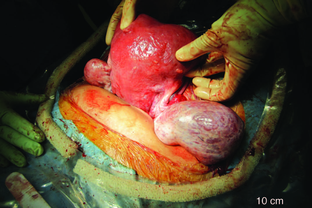
A 33 yr old woman presents with irregular menstrual bleeding and on examination has bilateral adnexal masses. At this original presentation, she was unexpectedly pregnant in the first trimester. Throughout her pregnancy these adnexal masses were presumed to be benign ovarian dermoids. However, at caesarean section the appearances were suspicious. Histological studies confirmed the presence of bilateral ovarian mature teratomas but also of a mucinous intestinal borderline tumour. A literature review of the management of adnexal masses in pregnancy is included.

Introduction

The diagnosis of adnexal masses during pregnancy has become increasingly common due to the widespread use of routine antenatal ultrasounds. [1-4,22] Despite the advancements in ultrasound technology, incidental adnexal masses are still being identified during caesarean section. [1,5,6]  Although the management of adnexal masses may differ if diagnosed during pregnancy or at caesarean section, the diagnostic limitations of antenatal ultrasonography may result in modifications in intraoperative or postpartum management.

Case Study

A 33 year old nulliparous woman presented to her general practitioner with concerns about irregular menstrual cycles that she had been experiencing since menarche at 13 years of age. The cycle duration was reported to be 30-90 days in length with a bleeding duration of approximately 7 days. The bleeding was described as “not heavy”.  She was in a long-term relationship and not using any form of contraception. She denied any pelvic or abdominal pain and had no gastrointestinal or genitourinary symptoms. She also denied any abnormal vaginal discharges or previous history of sexually transmitted diseases. Her cervical smears were up to date and had been consistently reported as “normal”. She had no past medical or surgical history but a strong family history of type 2 diabetes mellitus.

On examination, she appeared well and comfortable. Her abdomen was soft and non-tender. Speculum examination did not show any cervical abnormalities, however, on bimanual examination bilateral non-tender adnexal masses were found. A positive urine pregnancy was also detected.

A pelvic ultrasound demonstrated a bicornuate arcuate uterus and bilateral adnexal masses. The right adnexal mass measured 41x32x32mm and the left measured 72x59x64mm. Both masses were described as being echogenic, with no shadowing and no evidence of increased vascularity. The report suggested that the features exhibited were of bilateral ovarian mature teratomas (ovarian dermoids). Due to this, tumour markers, such as CA-125, were not performed. She was approximately 12 weeks pregnant at the time of the scan.

Throughout the pregnancy, pelvic ultrasounds were performed. These demonstrated an increase in the size of both adnexal masses. By 31 weeks of pregnancy the right ovary measured 140x110x65mm and the left ovary measured 77x52x55mm. No further ovarian changes were identified in subsequent ultrasounds up to the delivery date.

During the pregnancy the patient developed gestational diabetes mellitus that was poorly controlled resulting in a lower segment caesarean section being performed at 36 weeks of gestation. The preoperative plan was to perform a bilateral cystectomy at the time of the procedure.  This was based on the patient’s age, parity and the assumed benign nature of the ovarian masses.

Intraoperatively, a bicornuate arcuate uterus was identified with bilaterally enlarged ovaries (Figure 1). A bilateral cystectomy was performed without spillage of their contents, sparing the remaining ovarian tissue. Peritoneal washings were also taken for cytology based on the intraoperative findings of an unusual surface appearance of the right ovary (Figure 1). Following the operation, both cysts were bisected (Figure 2). The left cyst had typical dermoid features of sebaceous material and hair (Figure 3a). However, the right cyst had additional unusual features predominately of multiple mucinous cysts (Figure 3b).  Both cysts were sent for histopathology. The post-operative period was uneventful and she was discharged on day four.

28

Figure 1. Macroscopic appearance of the bicornuate arcuate uterus and bilaterally enlarged ovaries.


29

Figure 2. Macroscopic appearance of the left (L) and right (R) cysts following cystectomy.


30

Figure 3a. Bisected left cyst showing typical dermoid features.


31

Figure 3b. Bisected right cyst showing features of a dermoid cyst (D) and multi-loculated cysts (*). The line demarcates the dermoid-type tissue inferiorly and the mucinous component superiorly.

The histopathology report confirmed the left cyst to be a benign mature teratoma. However, the right cyst was reported as a mature teratoma together with a mucinous intestinal borderline tumour component. The peritoneal cytology did not reveal any evidence of malignant cells.

Discussion

Adnexal masses are detected in approximately 1-4% of all pregnancies. [2,9,10,24]  In most cases, the adnexal mass is diagnosed incidentally on routine antenatal ultrasounds in an otherwise asymptomatic patient. [2,8-10] However, in some cases the patient can present with symptoms such as pain and/or signs of a palpable mass. [2] In this case study, the patient did not present with any abdominal pain; however on examination bilateral adnexal masses were palpable which were also confirmed on ultrasound scan.

The majority of adnexal masses are of ovarian origin, [2,9] however others may arise from extra-ovarian structures such as the fallopian tubes, uterus and non-gynaecological tissues (Table 1). Frequently encountered ovarian masses in pregnant women include: mature cystic teratomas, cystadenomas and functional cysts. [5,6] In most cases, the adnexal pathologies are unilateral. [3,5,7] In cases with bilateral masses, the most frequent diagnosis is ovarian cystic teratomas. [5] In this case study, the preoperative ultrasound reports suggested that the bilateral masses were also ovarian teratomas.

32

The management of adnexal masses detected during pregnancy is controversial. Both expectant and surgical approaches, each carrying specific risks and benefits, are possible. Factors including size, gestational age and sonographic appearance may influence the final management. [7,9]Ultrasonography is the imaging modality of choice for detecting adnexal masses in both pregnant and non-pregnant women. [4,11] However, ultrasonography has limitations and not all adnexal masses during pregnancy are detectable as the gravid uterus may obscure the visualisation and detection of such masses. [1,5] Although ultrasonography may assist in differentiating benign from malignant adnexal masses, [4,10,18,23] it is not useful in differentiating between benign and low malignant potential tumours preoperatively. [17] This was highlighted in this case report, whereby the right cyst was reported as showing features suggestive of a benign mature teratoma and therefore, a cystectomy was performed. Subsequent histological analysis provided a diagnosis of a borderline tumour, where a salpingo-oophorectomy may have been the procedure of choice.

Most adnexal masses detected during pregnancy will resolve spontaneously or significantly reduce in size without any interventions. [2,8-11,15] Furthermore, only 2-8% of adnexal masses are malignant. [7,22] Therefore most masses during pregnancy can be managed expectantly, [8,9,15] particularly if they are asymptomatic, less than 5-6cm in diameter, have a simple sonographic appearance and are diagnosed before 16 weeks of gestation. [8] However, serial observations and ultrasound scans throughout the pregnancy are recommended to monitor potential changes in these masses. [15] The masses may then be managed surgically at caesarean section or have repeat imaging performed 6-8 weeks post-partum in the case of vaginal deliveries. [15] However, some of the risks associated with this conservative approach include: torsion, cyst rupture, infection, obstructed labour or a delayed diagnosis of malignancy. [1,3,8,12,25-29]

By contrast, a surgical approach is indicated for those patients with adnexal masses during pregnancy that are symptomatic, greater than 5-6cm in diameter, have a complex sonographic appearance suggesting malignancy, or persist into the second trimester. [2,8] The ideal window for surgical intervention in pregnant women is in the early-mid second trimester, to reduce the risk of complications. [10,13] These complications include: spontaneous miscarriage, spontaneous rupture of the membranes, preterm labour, preterm birth and intrauterine growth restriction. [2,10,13,14] However, it is unknown whether these complications are due to the effect of surgery or anaesthesia. Although laparoscopy or laparotomy may be performed, particularly in the second trimester, laparoscopic surgery is considered more beneficial to the mother with few studies suggesting an effect on the developing fetus. [20,21]

The impact of adnexal masses on the developing fetus is largely affected by the natural history of the adnexal mass. Most adnexal masses within Table 1, in-and-of-themselves will not directly affect fetal development. However, iatrogenic surgical procedures, whether as prophylactic or reactive interventions, may result in miscarriage or premature labour. Rarely, is premature delivery indicated to deal with the adnexal mass. However, in the term pregnancy, a caesarean section may be possible at the same time.

Generally, corticosteroids and tocolytics are not administered prophylactically for surgical procedures dealing with adnexal masses in pregnancies greater than 24 weeks of gestation. They may, however, be indicated following surgery. [30]

For those adnexal masses that are detected incidentally during caesarean section, it is recommended that they be removed. [1,4] The recommended procedure is a cystectomy, whilst oophorectomy and salpingo-oophorectomy procedures may also be considered. [16] The rationale is to exclude the possibility of malignancy and to avoid the need for further surgical procedures following the caesarean section. [1,5,16]

In this case report, the adnexal masses were managed with an expectant approach, despite their increasing sizes and persistent nature throughout the pregnancy. A bilateral cystectomy was performed at caesarean section on the assumption that the masses were teratomas, which are predominately benign tumours. [19] However, the histological diagnosis differed and revealed a tumour with borderline malignant potential. A different management approach may have been taken if this was suspected during the pregnancy.

This case report demonstrates the diagnostic limitations of ultrasonography and the potential dependence on this modality in the management of adnexal masses in pregnancy.

Summary Points

  1. The diagnosis of adnexal masses during pregnancy has increased due to the widespread use of ultrasonography.
  2. Although ultrasonography is useful in diagnosing adnexal masses, there are limitations.
  3. The management of adnexal masses during pregnancy is controversial. There are two approaches, including an expectant and a surgical approach.
  4. The recommended management approach for incidental masses detected at caesarean section is extirpation.

Consent declaration

Consent  to  publish  this  case  report  (including  photographs)  was obtained from the patient.

Acknowledgements

We would like to acknowledge the University of Wollongong’s Graduate School of Medicine for the opportunity to undertake a selective rotation in the Department of Obstetrics and at The Wollongong Hospital during medical school. In addition, we would like to thank The Wollongong Hospital library staff (Christine Monnie, Sharon Hay and Gnana Segar) for their excellent assistance with the literature search for this publication.

Conflict of interest

None declared.

Correspondence

M Petinga: michael.petinga@sesiahs.health.nsw.gov.au

References

[1] Dede M, Yenen MC, Yilmaz A, Goktolga U, Baser I. Treatment of incidental adnexal masses at cesarean section: a retrospective study. Int J Gynecol Cancer. 2007;17(2):339-41.

[2] Graham GM. Adnexal Masses in Pregnancy: Diagnosis and Management. Donald School Journal of Ultrasound in Obstetrics and Gynecology. 2007;1(4):66-74.

[3] Kondi-Pafiti A, Grigoriadis C, Iavazzo C, Papakonstantinou E, Liapis A, Hassiakos D. Clinicopathological characteristics of adnexal lesions diagnosed during pregnancy or cesarean section. Clin Exp Obstet Gynecol. 2012;XXXIX(4):458-61.

[4] Li X, Yang X. Ovarian Malignancies Incidentally Diagnosed During Cesarean Section: Analysis of 13 Cases. Am J Med Sci. 2011;341(3):181-4.

[5] Ulker V, Gedikbasi A, Numanoglu C, Saygi S, Aslan H, Gulkilik A. Incidental adnexal masses at cesarean section and review of literature. J Obstet Gynaecol Res. 2010;36(3):502-5.

[6] Ustunyurt E, Ustunyurt BO, Iskender TC, Bilge U. Incidental adnexal masses removed at caesarean section. Int J Gynaecol Obstet. 2007;96(1):33-4.

[7] Balci O, Gezginc K, Karatayli R, Acar A, Celik C, Colakoglu M. Management and outcomes of adnexal masses during pregnancy: A 6-year experience. J Obstet Gynaecol Res. 2008;34(4):524-8.

[8] Hoover K, Jenkins TR. Evaluation and management of adnexal mass in pregnancy. Am J Obstet Gynecol. 2011;205(2):97-102.

[9] Spencer CP, Robarts PJ. Management of adnexal masses in pregnancy. The Obstetrician & Gynaecologist. 2006;8(1):14-9.

[10] Elhalwagy H. Management of ovarian masses in pregnancy. Trends in Urology, Gynaecology and Sexual Health. 2009;14(1):14-8.

[11] Zanetta G, Mariani E, Lissoni A, Ceruti P, Trio D, Strobelt N et al. A prospective study of the role of ultrasound in the management of adnexal masses in pregnancy. BJOG. 2003;110(6):578-83.

[12] Yacobozzi M, Nguyen D, Rakita D. Adnexal Masses in Pregnancy. Seminars in Ultrasound, CT and MRI. 2012;33(1):55-64.

[13] Hoffman MS, Sayer RA. Adnexal masses in pregnancy. OBG Management. 2007;19:27-44.

[14] Mazze RI, Kallen B. Reproductive Outcome After Anesthesia and Operation During Pregnancy: A Registry Study of 5405 Cases. Am J Obstet Gynecol. 1989;161(5):1178-85.

[15] Nick AM, Schmeler K. Adnexal Masses in Pregnancy. Perinatology. 2010;2:13-9.

[16] Cristian F, Gheorghe C, Marius C, Gheorghe F. Management of adnexal masses during cesarean section – advantages of exteriorizing the uterus technique seen in a twelve year retrospective study. Paper Presented at: WSEAS; 2012 July 14-17; Kos Island, Greece.

[17] Whitecar MP, Turner S, Higby MK. Adnexal masses in pregnancy: A review of 130 cases undergoing surgical management. Am J Obstet Gynecol. 1999;181(1):19-24.

[18] Alcazar JL, Merce LT, Laparte C, Jurado M, Lopez-Garcia G. A new scoring system to differentiate benign from malignant adnexal masses. Am J Obstet Gynecol. 2003;188(3):685-92.

[19] Reed N, Millan D, Verheijen R, Castiglione M. Non-epithelial ovarian cancer: ESMO Clinical Practice Guidelines for diagnosis, treatment and follow-up. Annals of Oncology. 2010;21(5):31-6.

[20] Guidelines Committee of the Society of American Gastrointestinal and Endoscopic Surgeons. Yumi H. Guidelines for diagnosis, treatment, and use of laparoscopy for surgical problems during pregnancy: this statement was reviewed and approved by the Board of Governors of the Society of American Gastrointestinal and Endoscopic Surgeons (SAGES), September 2007. It was prepared by the SAGES Guidelines Committee. Surgical Endoscopy. 2008;22(4):849-61.

[21] Koo Y, Kim HJ, Lim K, Lee I, Lee K, Shim J et al. Laparotomy versus laparoscopy for the treatment of adnexal masses during pregnancy. Aust N Z J Obstet Gynaecol. 2012;52(1):34-8.

[22] Bernhard LM, Klebba P, Gray D, Mutch D. Predictors of Persistence of Adnexal Masses in Pregnancy. Obstetrics & Gynecology. 1999;93(4):585-9.

[23] Chiang G, Levine, D. Imaging of Adnexal Masses in Pregnancy. J Ultrasound Med. 2004;23(6):805-19.

[24] Meissnitzer M, Forstner R. Adnexal Masses in Pregnancy. In: Hamm B, Ros PR, editors. Abdominal Imaging. Berlin: Springer-Verlag; 2013.

[25] Erdemoglu M, Kuyumcuoglu U, Guzel A. Clinical experience of adnexal torsion: evaluation of 143 cases. J Exp Ther Oncol. 2011;9(3):171-4.

[26] De Santis M, Licameli A, Spagnuolo T, Scambia G. Laparoscopic Management of a Large, Twisted, Ovarian Dermoid Cyst During Pregnancy – A Case Report. J Reprod Med. 2013;58(5):271-6.

[27] Apostolakis-Kyrus K, Indermaur M, Prieto J. Teratoma in Pregnancy – A Case Report. J Reprod Med. 2013;58(9):458-60.

[28] Ansell J, Bolton L. Spontaneous rupture of an ovarian teratoma discovered during an emergency caesarean section. Journal of Obstetrics & Gynaecology. 2006;26(6):574-5.

[29] Giuntoli R, Vang R, Bristow R. Evaluation and management of adnexal masses during pregnancy.Clin Obstet Gynecol.2006;49(3):492-505.

[30] James D, Steer P, Weiner C, Gonik B. High Risk Pregnancy: Management Options. Philadelphia: Elsevier/Saunders; 2006.

Categories
Original Research Articles

Clavicle fractures: An audit of current management practices at a tertiary hospital, a review of the literature and advice for junior staff

Background: The clavicle is one of the most commonly fractured bones in the body. Interns are often delegated to treat these cases in an emergency department. This audit looks at the adherence to a tertiary hospital’s clavicle fracture protocol and reviews the literature to provide suggestions on updates based on current evidence. Methods: A retrospective case note and radiograph audit was undertaken to assess adherence to current protocols for the calendar years 2012 and 2013. A literature search was performed to find the most up to date evidence for future clavicle fracture management. Results: There were 131 clavicle fractures reviewed. An AP x-ray was taken in 120/122 cases (98.3%). The Orthopaedic registrar was notified for 6/7 (86%) cases with respiratory, neurovascular or skin compromise. Up to 83/131 (63%) patients were provided with a broad arm sling. Mean initial follow up was at ten days (3-20 days) and 39/95 (41%) followed x-ray protocol at this review. Appropriate rehabilitation advice was documented in 12/82 (14.6%) cases and the mean duration until discharge was 52.25 days. Conclusion: Despite the high frequency of clavicle fractures there are still significant errors that can be, and are being, made in their management. It is important for all medical students and junior doctors to become familiar with this Orthopaedic condition, as it is a common presentation that is often initially managed by junior medical staff.

Introduction33

Clavicle fractures are one of the most common fractures in the adult with an annual incidence of 29-64 per 100,000 people, per year. [1] Fractured clavicles account for up to 5% of all fractures and up to 44% of fractures to the shoulder girdle. [1,2]

Clavicle fractures are commonly managed by junior staff, and the current adult fracture protocol at our institution guides this management (Figure 1). The protocol was issued in July 2006 and reviewed in 2009. However, this protocol remains based on evidence the most recent of which was published in 1997. [3–8] There has been an influx of published literature on clavicle fractures over the last decade providing more recommendations for which an updated protocol can be based, including two well-designed multi-centre randomised controlled trials. [1,9,10,11,12,13] These articles demonstrate the shift from conservative management to surgical management for displaced and comminuted fractures of the adult clavicle.

This retrospective case note and radiograph audit firstly assesses the adherence of management practices at a tertiary hospital to the current institutional protocol for the calendar year of 2012 and 2013. Secondly, it discusses the standards of best practice in the current literature, with the view to providing recommendations for alterations to hospital protocol and management practice.

Methods

Process

In preparing for this audit a literature review was conducted to identify the gold standard and best practice guidelines for the investigation, management, and rehabilitation of clavicle fractures. Additionally, the hospital intranet was searched for any further documents including protocols, information sheets, and patient handouts. Consultation with the physiotherapy (PT) and occupational therapy (OT) departments was undertaken to assess any current gold standards, best practice or unwritten guidelines.

The case notes and radiographs of patients identified with a clavicle fracture were reviewed and adherence to the current protocol was assessed. Specifically, adherence to the following aspects of the protocol was scrutinised (Figure 1).

  1. All patients are to receive an anterior to posterior (AP) x-ray
  2. The orthopaedic registrar must be notified if there is respiratory, neurovascular or overlying skin compromise
  3. All patients are to receive a broad arm sling for acute management
  4. Outpatient follow up is to be booked for two weeks post injury
  5. Only postoperative patients are to have an x-ray on arrival (XROA) at the two week follow up
  6. All patients are to begin pendulum exercises immediately, range of motion (ROM) from two weeks, full active ROM (AROM) from 6 weeks or after clinically healed
  7. Return to sport (RTS) should be delayed for at least 4-6 months.

34

Figure 1. The current adult clavicle fracture protocol for which adherence was audited.

Ethical approval

This audit was reviewed and approved by the local clinical human research ethics committee. No identifiable patient data was collected and all records were viewed on site in the medical records department. A retrospective case note and electronic record audit was performed for the calendar year of 2013. As insufficient data was available to make reliable conclusions an additional calendar year, 2012, was included.

Patient recruitment

With the assistance of the orthopaedic department and the support of the project manager, all patients with clavicle fractures who presented to the emergency department (ED) or who were admitted to the wards in the calendar years 2012-13 were included. The hospital coding system, Inpatient Separations Information System (ISIS), was searched using the World Health Organisation (WHO) International Classification of Diseases, version ten, (ICD10) codes for all clavicle fracture admissions, S4200-3 inclusive. In addition the ED database for the two calendar years was hand-searched for provisional diagnoses relevant to clavicle fractures. This limited selection bias caused by spelling errors if searched electronically. These searches provided a list of 141 patient unit record numbers (URN) that were provided to medical records for retrieval.

Data retrieval

All case notes were reviewed immediately once available to minimise loss to the removal of records. Missing records were re-requested and viewed on multiple occasions until all records had been accounted for. Despite multiple searches two case notes were unable to be retrieved, being listed on the system as in stock but unable to be located by staff. For these two cases the electronic records were viewed to minimise selection bias and to ensure all patients were analysed.

Each patient file was meticulously studied and cross-referenced against the electronic discharge summaries and encounters, in addition to radiological analysis using a picture archiving and communication system.

Data analysis

Data was collected and stored in a Microsoft Excel (Copyright Microsoft Corporation 2010) spread sheet. Simple descriptive statistical analysis was performed using IBM SPSS version 22 (Copyright IBM Corporation and other(s) 1989, 2013).

Standards for adherence

In consultation with the orthopaedic department it was determined that 90% compliance with the current protocol would be deemed acceptable. Adherence was analysed collectively for the entire cohort but also separately for the two calendar years, surgical versus non-surgical patients, and then again against current literature recommendations. Only the collective data will be presented.

Results

Recruitment and demographics

The database searches resulted in the retrieval of 141 patient URNs. Of these 131 were new clavicle fractures. There were 99 males and 42 females of which 47 fractures were on the right and 84 on the left (Table 1). The dominant arm was affected in 17 cases, non-dominant in 34, and not documented in 80 cases. There were three medial (2.3%), 93 central (70.1%) (Figure 2), 34 lateral (25.9%), and one both middle and lateral (other 0.8%). This is almost identical to the fracture pattern distribution reported by Robinson. [11] Associated injuries were documented in 34/131 (26%) cases. This is slightly less than the 36% reported by Nowak, Mallmin & Larsson however there are differences between the skin abrasions that were included in each study. [14]

35


Radiological adherence

An AP radiograph was taken for 120/122 (98.3%) patients (nine outside films). The two patients that did not have an x-ray both re-presented and subsequently had an x-ray identifying a clavicle fracture and were therefore included in this audit. Only 97/122 (79.5%) had two views taken of their clavicle fracture on presentation despite current literature and orthopaedic dogma dictating that every fracture should be viewed from two angles and include two joints. [15,16]

At the two week review 39/95 (41%) cases followed protocol regarding XROA. The conservative group were not required to have an x-ray at two weeks with adherence in 30/85 (35.3%), however, 13 of these had x-rays at subsequent appointments. For the conservative cases that did have x-rays at their first outpatient department (OPD) appointment 3/55 (5.5%) resulted in a change of management toward surgery. Nine out of ten (90%) surgical patients had an x-ray to check the position of the metalwork at two weeks as required.

36

Figure 2. The typical middle third clavicle fracture that presents a management dilemma.

Broad arm sling

Compliance with the protocol regarding the broad arm sling application is summarised in Figure 3. Of note 6/32 (18.8%) patients who were provided with a collar and cuff for acute management showed progressive displacement and five of these required surgical fixation. If the benefit of the doubt is given and all those who were documented as given a ‘sling’ are combined with the BAS then 83/131 (63.3%) patients were correctly treated.

37

Figure 3. Illustrating the adherence of staff to provide a broad arm sling as first line management. BAS = Broad Arm Sling, C+C = Collar and Cuff, #HOH = Fractured Head of Humerus, OPD = Outpatient Department.

Registrar notification

The orthopaedic registrar was notified 46 times for the 131 cases analysed (35%), although according to the protocol they are only required to be notified if there is respiratory, neurovascular, or skin compromise. In this case they were notified on six out of seven occasions (86%). However, if they were also required to be notified for displaced fractures >20mm and shortened >15mm in addition to the associated injuries in the protocol, then they were notified in 27/51 (53%) cases. Cases of which the orthopaedic registrar was not informed of include one floating shoulder, three ACJ separations, one head of humerus (HOH) fracture, and one patient with ipsilateral rib fractures 1-5.

Prior to the outpatient follow up six patients re-presented to ED, two patients on multiple occasions. Of these two were treated with a collar and cuff and one with a sling.

Complications or associated injuries were present in 18/131 cases (13.7%), five with tented/compromised skin, six ACJ separations, four floating shoulders, one ipsilateral HOH fracture, one ulnar nerve paraesthesia, and one patient with multiple ipsilateral rib fractures. Of these six underwent surgical fixation.

There were 14 surgeries (11 middle third, three lateral) and eleven patients received private orthopaedic management (Table 2).

38

Rehabilitation

There were 82 patients followed up in the orthopaedic OPD clinic. Twelve of these (14.6%) had sufficient documentation to suggest the patient had been provided with appropriate rehabilitation advice. These included 4/9 (44%) surgical cases and 8/73 (11%) conservative cases. This left a large cohort of patients that had been given some or no advice on what rehabilitation they could perform. Six patients attended their six-week review having been immobilised in their sling for the entire duration leading to stiff painful shoulders. In eight case notes there is mention of seeking physiotherapy treatment of which two cases were treated by the hospitals physiotherapy department. There are no current physiotherapy handouts or protocols and the occupational therapy department has no involvement in the management or rehabilitation of clavicle fractures.

39Discussion

Clavicle fractures are a common presentation to any emergency department, representing 5% of all fractures, and are often managed by junior staff. This audit demonstrates that there is still some mismanagement and that further education of junior staff is required.

The gold standard for initial radiological review of clavicle fractures remains to be elucidated but current evidence agrees on the standard AP radiograph plus a second radiograph tilted on an angle. [15,16,17] Only 97/131 (74%) patients received two different views of their clavicle fracture on presentation. The angle of the second view ranged from 5-30 degrees AP cephalic tilt, with each five-degree increment in between. The optimal angle and direction of this second radiograph varies among authors with some recommending a posterior to anterior (PA) 15-degree cephalic tilt, while others recommend an AP 15-degree caudal tilt. [15,16,17] This makes it difficult to compare fracture patterns between clavicles when determining clinical management and also for future retrospective analysis. It may be preferable to have two views from the same side to limit the manoeuvring of patients and reduce the time demand on the radiological department. A second AP view with 20 degrees cephalic tilt has been recommended and would be the technically easiest view with minimal changes to current practices. [9]

Conservative treatment remains the management of choice for isolated non-displaced clavicle fractures. [12,18] The broad arm sling is recommended for clavicle fractures, [19] as the collar and cuff allows traction on the arm that risks further displacement of fracture segments. [20,21] The figure-of-eight slings have not been shown to be superior to the broad arm sling and are more uncomfortable and difficult to use. [22] Our results demonstrate that the collar and cuff is still being provided in many cases over the preferred broad arm sling. This may be due to confusion with the fractured neck of humerus that requires distracting forces for fracture alignment. One fifth of patients provided with collar and cuffs demonstrated significant progression of fracture displacement with many of these requiring surgical fixations.

While most isolated non-displaced fractures of the clavicle are managed conservatively, it is important to know when to refer to the orthopaedic department for review. Clear operative indications include compromise to the skin, nerves, vasculature, and grossly displaced/comminuted fractures. [23] Some authors also advocate for the primary fixation of clavicle fractures with multi-trauma, ipsilateral shoulder injuries, acromioclavicular joint (ACJ) involvement, high velocity mechanisms, or young active individuals. [24,25] For these patients, the surgeon’s preference continues to dictate treatment. [26] As such it is difficult to create a protocol to mandate management and the protocol is rather a guide to prompt management considerations.

Furthermore, over the past ten years there has been a growing body of literature increasing the relative and absolute surgical indications for clavicle fractures [13,23,24,27] Newer studies have highlighted that previously used outcomes may not be the best end points for assessing management. Previously, radiological union was the sole primary outcome for assessment of fracture management. [18,28] Recent studies focussing on patient-centred outcomes such as pain and functional capacity however have highlighted a disparity between radiological union and satisfactory objective and subjective patient-centred outcomes. [9-14] Furthermore, older studies have included children in outcome assessments, despite the greater regenerative capacity of this younger age group prior to fusion of the medial growth plate, leading to an over estimation of adult clavicle fracture recovery in these older studies. [20]

Recent studies have demonstrated sequelae including pain and neurological deficits occurring in up to 46% of clavicle fractures. [29] These sequelae are more likely in non- or mal-union of the fracture. Non-union rates for adult patients are as high as 15% and symptomatic mal-union up to 20-25%. [13] Risk factors for non-union include:

  • Smoking (33.3%)
  • Increasing Age and Female gender (often less symptomatic in this population)
  • Shortening >20 mm (increasing shortening increases non-union/mal-union)
  • Displacement >15 mm (27%)
  • Comminuted fractures (21.3%). [24,30]

The non-union rate for surgically treated patients’ is around 1-2%. [9,10,13] The number needed to treat (NNT) if all displaced clavicle fractures were operated on to prevent one symptomatic non-union would be 7.5, this number is reduced to 4.6 if symptomatic mal-unions are included. [13,24] The NNT drops to 1.7 if only those with multiple risk factors for non-union were surgically fixed. [24] Surgical risks must be considered however, including infection, implant irritation, neurological damage, and even death. [9,10,31] For widely displaced mid third clavicle fracture surgical plate fixation has been shown to be superior to conservative management. [9,10]

In this audit, the orthopaedic registrar was notified for most cases where there was neurovascular, respiratory, or skin compromise. However, in the current protocol there is mention of displacement and shortening with a decision to be made on whether the fracture is stable or unstable and whether the patient receives surgical or conservative management. If the orthopaedic registrar was required to be notified to make this decision then they were only notified in half the cases. It may be seen as an increased demand on the ED if they had patients waiting in beds for an orthopaedic review if it was considered unnecessary.

Murray and colleagues have shown that shortening and displacement are risk factors for non-union and it has been shown that delaying surgery results in worse functional outcomes. [24,32,33] Hence, it would be prudent to have an orthopaedic review in ED to make the clinical decision regarding management immediately. While this would increase the demand on orthopaedic staff to review x-rays and or patients from ED it also has the potential to decrease outpatient demand. By providing patients with the correct treatment immediately, early (< 2 weeks) outpatient follow up can be avoided and the six re-presentations to ED potentially avoided. Additionally if more accurate predictions are made for probable outcomes then work loads can be reduced by minimising the patients that are treated for extended periods or who fail conservative treatment and undergo additional surgical fixation.

There was a large variation in time to outpatient follow-up after ED discharge. This potentially reflects the uncertainty of the ED staff in their management of the patient. Rather than discharging the patient with appropriate sling, pain medication, and early rehabilitation advice they are requesting very early orthopaedic outpatient follow up essentially doubling the patient’s visits.

Radiological evaluation at the two-week review was over-utilised with correct use in less than half of the patients. This again could reflect uncertainty among medical staff about management. Of the 55 conservative patients undergoing multiple x-ray evaluations, a change in management was only initiated in three cases. Progressive displacement of the fracture ends has been recognised in one third of cases over a 5-year period by Plocher et al. who recommends serial x-rays for the first 3 weeks. [25] However, repeated x-rays are not useful unless the information is going to be used to guide clinical decision-making. Again, identifying patients at risk of progressive displacement (such as high velocity injuries) or those on the cusp of surgical intervention and providing early decision-making could remove the need to wait and watch.

Advice on rehabilitation was poorly documented and may reflect that it was not provided adequately in many cases. Documentation of education provided is an important part of keeping legal records and only one-fifth of patients had documented evidence that rehabilitation advice was provided. It was evident that no advice had been given for the six patients that returned to the six-week review having remained immobilised in the sling. Rehabilitation timeframes are largely based on expert opinion and are generally consistent with the current practices. [9,20,34] There has been limited research into the optimal timeframe to return to sport, however one study followed 30 patients after plate fixation and 20 had returned to sport after twelve weeks. [35] Three conservative patients re-fractured within 3-5 months in our audit population suggesting that returning to full contact sport should be delayed greater than six months.

The reliability of the results provided is limited by the dependence on multiple steps in accurately identifying and documenting clavicle fractures. These factors include:

  • Staff in ED diagnosing the clavicle fractures and documenting accordingly
  • Administration clerk in ED transcribing appropriate provisional diagnoses
  • Hospital coding officers applying the correct ICD10 codes
  • Human error is possible in misidentifying data in the notes
  • Human error in transcribing data into SPSS
  • Especially dependent on the accuracy of the doctor’s written case notes and discharge summaries for the above steps and also audit data collection.

Every effort was taken to ensure that the data collected was accurate including cross-referencing across multiple platforms. In stating this, however, the data collection for the variables dependent on documentation can only be as accurate as the written information and may not be a true representation of actual events. This would impact most significantly on the reporting of the sling provided, orthopaedic registrar notification, and rehabilitation advice given.

The methods used do not capture the cases that were directly referred to the outpatient department without first presenting to ED or as an inpatient. However, the majority of this audit involved looking at the initial management, as per the protocol, and therefore these cases were not required.

Conclusion

Clavicle fractures are a common presentation to any emergency department and are often managed by junior staff. This audit demonstrates that there is still some mismanagement, particularly in radiological assessment, sling prescription, and knowledge of protocol for registrar notification, outpatient follow-up and rehabilitation. Furthermore, new evidence indicates that the current protocol at this institution requires updating to clarify the requirements for referral and allow earlier interventions or rehabilitation. In summary, recommended radiological views are a standard AP and a second AP with 20 degrees cephalic tilt. Isolated non-displaced fractures of the clavicle are almost always managed conservatively, however, it is important to know when to refer to the orthopaedic department for review. This is always necessary if there are associated injuries or pending complications. It is also recommended that all displaced or comminuted fractures be referred for an orthopaedic opinion. The broad arm sling is the immobilisation technique of choice and not the collar and cuff for clavicle fractures because the collar and cuff allows distracting forces that risks further displacement of the fracture segments. Early rehabilitation is required to prevent painful stiff shoulders.

Acknowledgements

The authors would like to thank Katharina Denk, orthopaedic research assistant for her work in identifying patients records for inclusion in this audit.

Conflict of Interest

 None declared

Correspondence

D M George: daniel.george@health.sa.gov.au

Reference:

[1] Khan LA, Bradnock TJ, Scott C, Robinson CM. Fractures of the clavicle. J Bone Joint Surg Am. 2009;91(2):447–60.

[2] Postacchini F, Gumina S, De Santis P, Albo F. Epidemiology of clavicle fractures. J Shoulder Elb Surg. 2002;11(5):452–6.

[3] Andersen K, Jensen PO, Lauritzen J. Treatment of clavicular fractures. Acta Orthop Scand. 1987;58(1):71–4.

[4] Bostman O, Manninen M, Pihlajamaki H. Complications of plate fixation in fresh displaced midclavicular fractures. J Trauma Inj Infect Crit Care. 1997;43(5):778–83.

[5] Nordqvist A, Redlund-Johnell I, von Scheelel A, Petersson CJ. Shortening of clavicle after fracture: Incidence and clinical significance, a 5-year follow-up of 85 patients. Acta Orthop Scand. 1997;68(4):349–51.

[6] Hill JM, McGuire MH, Crosby LA. Closed treatment of displaced middle-third fractures of the clavicle gives poor results. J Bone Joint Surg Br. 1997;79-B(4):537–9.

[7] Davids PHP, Luitse JSK, Strating RP, van der Hart CP. Operative treatment for delayed union and nonunion of midshaft clavicular fractures: ao reconstruction plate fixation and early mobilization. J Trauma Inj Infect Crit Care. 1996;40(6):985–6.

[8] Hutchinson MR, Ahuja GS. Diagnosing and Treating Clavicle Injuries. Phys Sportsmed. 1996;24(3):26–36.

[9] Canadian Orthopaedic Trauma Society. Nonoperative treatment compared with plate fixation of displaced midshaft clavicular fractures. A multicenter, randomized clinical trial. J Bone Joint Surg Am. 2007;89(1):1–10.

[10] Robinson CM, Goudie EB, Murray IR, Jenkins PJ, Ahktar MA, Foster CJ, et al. Open reduction and plate fixation versus nonoperative treatment for displaced midshaft clavicular fractures: a multicentre, randomized, controlled trial. J Bone Joint Surg Am. 2013;95-A(17):1576–84.

[11] Robinson AM. Fractures of the clavicle in the adult epidemiology and classification. J Bone Joint Surg Br. 1998;80-B(3):476–84.

[12] Lenza M, Buchbinder R, Johnston R, Belloti J, Faloppa F. Surgical versus conservative interventions for treating fractures of the middle third of the clavicle (Review). Cochrane database Syst Rev Online. 2013;(6):CD009363.

[13] Mckee RC, Whelan DB, Schemitsch EH, Mckee MD. Operative versus nonoperative care of displaced midshaft clavicular fractures: a meta-analysis of randomized clinical trials. J Bone Joint Surg Am. 2012;94-A(8):675–84.

[14] Nowak J, Mallmin H, Larsson S. The aetiology and epidemiology of clavicular fractures. A prospective study during a two-year period in Uppsala, Sweden. Injury. 2000;31(5):353–8.

[15] Axelrod D, Safran O, Axelrod T, Whyne C, Lubovsky O. Fractures of the clavicle: which x-ray projection provides the greatest accuracy in determining displacement of the fragments? J Orthop Trauma. 2013;3(electronic):1–3.

[16] Sharr JR, Mohammed KD. Optimizing the radiographic technique in clavicular fractures. J Shoulder Elbow Surg. 2003;12(2):170–2.

[17] Smekal V, Deml C, Irenberger A, Niederwanger C, Lutz M, Blauth M, et al. Length Determination in Midshaft Clavicle Fractures: Validation of Measurement. J Orthop Trauma. 2008;22(7):458–62.

[18] Neer C. Nonunion of the clavicle. J Amercan Med Assoc. 1960;172:1006–11.

[19] Lenza M, Belloti JC, Andriolo RB, Gomes Dos Santos JB, Faloppa F. Conservative interventions for treating middle third clavicle fractures in adolescents and adults. Cochrane database Syst Rev Online. 2009;2(2):CD007121.

[20] Jeray KJ. Acute midshaft clavicular fracture. J Am Acad Orthop Surg. 2007;15(4):239–48.

[21] Mckee MD, Wild LM, Schemitsch EH. Midshaft malunions of the clavicle. J Bone Joint Surg Am. 2003;85-A(5):790–7.

[22] McKee MD. Clavicle fractures in 2010: sling/swathe or open reduction and internal fixation? Orthop Clin North Am. 2010;41(2):225–31.

[23] Lenza M, Belloti JC, Gomes Dos Santos JB, Matsumoto MH, Faloppa F. Surgical interventions for treating acute fractures or non-union of the middle third of the clavicle. Cochrane database Syst Rev Online. 2009;(4):CD007428.

[24] Murray IR, Foster CJ, Eros A, Robinson CM. Risk factors for nonunion after nonoperative treatment of displaced midshaft fractures of the clavicle. J Bone Joint Surg Am. 2013;95(13):1153–8.

[25] Plocher EK, Anavian J, Vang S, Cole PA. Progressive displacement of clavicular fractures in the early postinjury period. J Trauma. 2011;70(5):1263–7.

[26] Heuer HJ, Boykin RE, Petit CJ, Hardt J, Millett PJ. Decision-making in the treatment of diaphyseal clavicle fractures : is there agreement among surgeons ? Results of a survey on surgeons ’ treatment preferences. J Shoulder Elb Surg. 2014;23(2):e23–e33.

[27] Zlowodzki M, Zelle BA, Cole PA, Jeray K, McKee MD. Treatment of acute midshaft clavicle fractures: systematic review of 2144 fractures. J Orthop Trauma. 2005;19(7):504–7.

[28] Rowe CR. An atlas of anatomy and treatment of midclavicular fractures. Clin. Orthop. Relat. Res. 1968;58:29–42.

[29] Nowak J, Holgersson M, Larsson S. Can we predict long-term sequelae after fractures of the clavicle based on initial findings? A prospective study with nine to ten years of follow-up. J Shoulder Elb Surg. 2004;13(5):479–86.

[30] Robinson CM, Court-Brown CM, McQueen MM, Wakefield AE. Estimating the risk of nonunion following nonoperative treatment of a clavicular fracture. J Bone Joint Surg Am. 2004;86-A(7):1359–65.

[31] Wijdicks FJ, Van der Meijden OA, Millett PJ, Verleisdonk EJ, Houwert RM. Systematic review of the complications of plate fixation of clavicle fractures. Arch Orthop Trauma Surg. 2012;132(5):617–25.

[32] Potter JM, Jones C, Wild LM, Schemitsch EH, McKee MD. Does delay matter? The restoration of objectively measured shoulder strength and patient-oriented outcome after immediate fixation versus delayed reconstruction of displaced midshaft fractures of the clavicle. J Shoulder Elb Surg. 2007;16(5):514–8.

[33] George DM, McKay BP, Jaarsma RL. The long-term outcome of displaced mid-third clavicle fractures on scapular and shoulder function: variations between immediate surgery, delayed surgery, and nonsurgical management. J Shoulder Elb Surg. 2015; 24(5):669-76.

[34] Kim W, McKee MD. Management of acute clavicle fractures. Orthop Clin North Am. 2008;39(4):491–505.

[35] Meisterling SW, Cain EL, Fleisig GS, Hartzell JL, Dugas JR. Return to athletic activity after plate fixation of displaced midshaft clavicle fractures. Am J Sports Med. 2013;41(11):2632–6.

Categories
Feature Articles

Epidural analgesia during labour: Friend or foe? A reflection on medicine, midwives and Miranda Kerr

Choosing a method of pain relief for childbirth is an extremely personal, and often well-considered, decision. For many women, childbirth is the most painful experience they will ever encounter. It is no surprise that a number of pharmacological and non-pharmacological methods have been developed to help manage this painful and sometimes traumatic experience. In Western cultures, epidural analgesia (EA), as well as a number of other methods, is widely used, and its benefits (and risks) are well documented. [1-3] Despite the generally positive evidence base, many women choose not to use EA during their labour. [1, 4, 5] Clearly, there are other factors that influence their choice of pain relief (or lack thereof). Personal attitudes towards the acceptability of labour pain and fear of the process are key, but outside influences can be significant. [1, 5, 6] Those around her – her doctors and/or midwives, her family and friends – will almost certainly have shaped her attitude. However, public pressure generated by celebrities such as Miranda Kerr may influence a woman’s decision more than we realise. In the age of social media, where opinions are abundant and conflicting, women may be more confused than ever: is an epidural a friend or a foe?

There are a number of methods available for managing pain during the labour process. In discussing these options it often becomes a balancing act between what the woman considers to be an acceptable level of pain, with an acceptable level of risk – a highly personal decision that relies on a woman being able to adequately understand the risks and consequences. Options for analgesia may be non-pharmacological, such as massage, breathing exercises and transcutaneous electrical nerve stimulation (TENS), which have limited evidence of efficacy but appear to improve satisfaction with the childbirth experience (compared to placebo). [2, 3] Pharmacological choices include:

  • Inhalation agents (i.e. nitrous oxide), which relieve pain compared with placebo but are associated with nausea, vomiting and dizziness [2, 3]
  • Systemic opioids, which are less effective than regional analgesics and frequently cause nausea and sedation [2, 7]
  • Local anaesthetic nerve blocks, which are especially useful for instrumental delivery and episiotomy (often in conjunction with EA) [8]
  • Regional analgesia, including EA, spinal anaesthesia, and combined spinal-epidural anaesthesia (CSE)

EA is widely used for pain relief in labour and involves injection of a local anaesthetic (such as bupivacaine) into the epidural space. [2] It is typically given with an opioid such as fentanyl to limit the amount of local anaesthetic required for efficacy. This also allows the woman greater ability to bear down and push during the second stage of labour. EA effectively relieves pain (compared to opioids or placebo) but does increase the risk of instrumental delivery and caesarean section for fetal distress, and may prolong the second stage of labour by up to two hours. [2, 3, 9, 10] Other potential adverse effects include hypotension, motor blockade, fever and urinary retention (requiring an indwelling urinary catheter). [3, 7] Fear of EA side effects has been noted as a key predictor as to whether a woman will elect for EA, with one study suggesting fear of EA side effects decreases EA uptake by half. [1] As EA allows insertion of a catheter, the medication can be given by bolus injection, continuous infusion or via a patient-controlled pump. This is in contrast to spinal anaesthesia (injection of local anaesthetic into the subarachnoid space), which, while faster and safer, does not allow insertion of a catheter for continuing analgesia. [2, 10] In many centres, a combined spinal and epidural anaesthetic (CSE) is given, where a single injection of local anaesthetic is inserted into the subarachnoid space (for fast onset of pain relief) in addition
to insertion of an epidural catheter for ongoing pain management. [10]

Women have widely differing views on what level of pain should be expected when giving birth. Evidence suggests that women who are more fearful of labour pain have a higher likelihood of choosing elective caesarean, and if they do choose labour, a higher chance of having an epidural. [1, 6] In contrast, women who are more accepting of labour pain, and more confident in their ability to cope with it, are generally more likely to decide against EA. [1, 6, 11] Other personal factors that increase the likelihood of a woman choosing EA include having a previous EA, partner preference, and attending a childbirth class. [4, 11] In addition, the attitudes and experiences of family and friends can influence a woman’s decision. It has been shown that women with friends or family who have had positive experiences with EA are more likely to choose EA themselves. [1] Likewise, hearing stories about how excruciatingly painful childbirth is may increase anxiety about the pain and increase EA uptake for primiparous women. [1]

Looking beyond a woman’s immediate circle of family and friends reveals another potential influence – celebrities and the media. There appears to be a widespread opinion (particularly amongst celebrities) that birth should be “natural”, which presumably refers to a lack of intervention. [12] Just as organic, gluten-free, paleo, and #cleaneating have taken off, a similar trend appears to be on the rise in childbirth. Perhaps next we will see the emergence of “organic” labour wards. Miranda Kerr had the media buzzing following her comments about having “a natural birth without pain relief” and not wanting a “drugged-up baby.” [13, 14] Whilst it was absolutely her choice to give birth “naturally” and opt out of pain medication, her celebrity status mean that her personal experiences and opinions are likely to influence the behaviours and attitudes of women all over Australia (and potentially the world). By going out of her way to state in her official announcement of the birth of her son: “I gave birth to him naturally; without any pain medication” it infers that those who decide otherwise are making the ‘wrong’ decision. [13, 14] Sweeping declarations like this have the potential to be damaging to women who did elect to use EA or needed a caesarean section. It may be that public assertions about their choices, made by Miranda Kerr and other celebrities such as Teresa Palmar and Gisele Bündchen, have turned EA into the enemy. [12] Such statements generate significant media interest and controversy, and have led to the emergence of the term “the smug natural birth” as well as suggestions that giving birth has become “a competitive sport.”[12]

But it’s not just celebrities and models that have a problem with epidurals. There is a difference of opinion between midwives and obstetricians as to how often epidural analgesia should be used. [15] An article published in Midwifery Today in 2010 referred to epidurals as “the drug trip of the current generation”, and even compared anaesthetists to “street drug pushers.” [16] Whilst clearly
this does not represent the views of all midwives, it is concerning that a prominent publication can present these opinions as if they were fact. This article also advised it’s audience of birth practitioners to remember that “a woman who can sit still long enough to have an epidural inserted during labor can have a relatively painless, unmedicated birth if she were provided adequate birth support in the home setting.”[16] This misinformation is dangerous given the fact that RANZCOG does not support the practice of planned homebirths due to its inherent and proven risks. [17] The reluctance of some midwives to offer EA has been well documented elsewhere. [15, 18, 19]

Furthermore, a number of Australian studies have found that the rate of epidural analgesia uptake is much higher in private hospital patients versus those seen in the public system. [20, 21] A New South Wales study from 2012 reported a 40% larger uptake of EA in private hospitals compared with public, as well as an overall increase in interventions. [20] This is similar to previous Australian data reporting a 50% increase in uptake of EA in private versus public care. [21] It is clear that many women are not in a position to choose whether they receive public or private care, but nonetheless it is apparent that where one gives birth has an impact on whether an EA will be performed or not. This raises issues of appropriate health care expenditure and a potential two-tier system in Australia that deserves adequate discussion and reflection in its own right. [20]

Ultimately, women should feel free to choose whatever pain relief they believe will help them most during labour, or to opt for none at all. Furthermore, whilst this reflection has focused primarily on women determining a birth plan in the antenatal period, women who choose non-pharmacological methods during that period should also feel free to progress to a pharmacological method during labour if they are not coping with the pain. It is important that women are informed and feel empowered to make these decisions, and this involves adequate discussion of the benefits and potential adverse effects of all their options. As the doctor – whether we are the obstetrician, the anaesthetist, the GP or perhaps even the resident, it is our job to ensure the patient fully understands that discussion. However, in order to communicate benefits and risks effectively we need an understanding of what influences a woman’s choice when it comes to pain medication, even more so when attempting to navigate the controversial minefield that is childbirth. Evidence-based medicine is brilliant, but sometimes we live in an evidence bubble – so influenced by statistics that we might forget to look outside at how the opinions and actions of others can also shape our patients’ decisions. To our patients, percentages may mean nothing in the face of Miranda Kerr and organic kale smoothies. A thorough discussion of a woman’s fears and attitudes towards the birthing process is undoubtedly a crucial component of comprehensive antenatal care.

Acknowledgements

None.

Conflict of interest

None declared.

Correspondence

C de Rooy: cderooy@outlook.com

Reference List:

[1] Van den Bussche E, Crombez G, Eccleston C, Sullivan M. Why women prefer epidural analgesia during childbirth: the role of beliefs about epidural analgesia and pain catastrophizing. European Journal of Pain. 2007;11(3):275-82.

[2] Anim-Somuah M, Smyth R, Jones L. Epidural versus non-epidural or no analgesia in labour (review). The Cochrane Database of Systematic Reviews. 2011.

[3] Jones L, Othman M, Dowswell T, Alfirevic Z, Gates S, Newburn M, et al. Pain management for women in labour: an overview of systematic reviews. The Cochrane Database of Systematic Reviews. 2012.

[4] Horowitz E, Yogev Y, Ben-Haroush A, Kaplan B. Women’s attitude towards analgesia during labour – a comparison between 1995 and 2001. European Journal of Obstetrics & Gynecology and Reproductive Biology. 2004;117:30-2.

[5] Petersen A, Penz S, Mechthild M. Women’s perception of the onset of labour and epidural analgesia: a prospective study. Midwifery. 2013;29(284-93).

[6] Haines H, Rubertsson C, Pallant J, Hildingsson I. The influence of women’s fear, attitudes and beliefs of childbirth on mode and experience of birth. BMC Pregnancy and Childbirth. 2012;12.

[7] Narayan R, Hyett J. Labour and delivery. In: Abbott J, Bowyer L, Finn M, editors. Obstetrics & Gynaecology: an evidence-based guide. NSW: Elsevier Australia; 2014.

[8] Schinkel N, Colbus L, Soltner C, Parot-Schinkel E, Naar L, Fournie A, et al. Perineal infiltration with lidocaine 1%, ropivacaine 0.75%, or placebo for episiotomy repair in parturients who received epidural labor analgesia: a double-blind randomized study. International Journal of Obstetric Anesthesia. 2010;19(3):293-97.

[9] Cheng Y, Shaffer B, Nicholson J, Caughley A. Second stage of labor and epidural use: a larger effect than previously suggested. Obstetrics and Gynaecology. 2014;123:527-35.

[10] Heesen M, van de Velde M, Klohr S, Lehberger J, Rossaint R, Straube S. Meta-analysis of the success if block following combined spinal-epidural vs epidural analgesia during labour. Anaesthesia. 2014;69(1):64-71.

[11] Harkins J, Carvalho B, Evers A, Mehta S, Riley E. Survey of the factors associated with a woman’s choice to have an epidural for labor analgesia. Anesthesiology Research and Practice. 2010.

[12] Wainwright H. Terrific, now birth has become a competitive sport. Mamamia [Internet]. 2014. Available from: http://www.mamamia.com.au/celebrities/natural-birth/.

[13] Mamamia Team. This is why Miranda Kerr had a natural birth. Mamamia [Internet]. 2012. Available from: http://www.mamamia.com.au/news/natural-birth-miranda-kerr/.

[14] Wellman V. ‘I didn’t want a drugged up baby’: Miranda Kerr on her decision not to have a epidural during son Flynn’s birth. The Daily Mail [Internet]. 2012. Available from: http://www.dailymail.co.uk/femail/article-2169435/Miranda-Kerr-decision-epidural-son-Flynns-birth.html.

[15] Reime B, Klein M, Kelly A, Duxbury N, Saxell L, Liston R, et al. Do maternity care provider groups have different attitudes towards birth? BJOG: an International Journal of Obstetrics and Gynaecology. 2004;111(12):1388-93.

[16] Cohain J. The epidural trip: why are so many women taking dangerous drugs during labour? Midwifery Today. 2010;65:21-4.

[17] The Royal Australian College of Obstetricians and Gynaecologists. RANZCOG Clinical Guidelines: Home births 2011. Available from: http://www.ranzcog.edu.au/college-statements-guidelines.html.

[18] Potera C. Evidence supports midwife-led care models: fewer premature births, epidurals, and episiotomies; greater patient satisfaction. The American Journal of Nursing. 2013;113:15.

[19] Raini R, Salamut W, Chowdhury P, Daborwska D. Epidurals: myths and old wives tales – what are the midwives telling our patients? Anaesthesia. 2013;68:98.

[20] Dahlen H, Tracy S, Tracy M, Bisits A, Brown C, Thornton C. Rates of obstetric intervention and associated perinatal mortality and morbidity among low-risk women giving birth in private and public hospitals in NSW (2000-2008): a linked data population-based cohort study. BMJ Open. 2014;2.

[21] Roberts C, Tracy S, Peat B. Rates for obstetric intervention among private and public patients in Australia: a population based descriptive study. British Medical Journal. 2000;321:137-41.